Download
1 / 19
190 likes | 548 Views
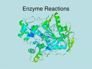
Hypersensitivity reactions: Antibody-mediated (type II) cytotoxic reactions and immune complex (type III) reactions. Type II. Tissue/cell damage due to direct action of antibody or complement Antibody dependent cell-mediated cytotoxicity (ADCC) Examples

E N D
Hypersensitivity reactions: Antibody-mediated (type II) cytotoxic reactions and immune complex (type III) reactions
Type II • Tissue/cell damage due to direct action of antibody or complement • Antibody dependent cell-mediated cytotoxicity (ADCC) • Examples • Transfusion reactions (Rhesus incompatibility) • Organ transplants • Autoimmune type II reactions - ‘cold’ agglutinins • Type II drug reactions - ‘penicillin’
Figure 15.1Schematic illustration of three different mechanisms of antibody-mediated injury in type II hypersensitivity. (A) Complement-dependent reactions that lead to lysis of cells or render them susceptible to phagocytosis. (B) Antibody-dependent cell-mediated cytotoxicity (ADCC). IgG-coated target cells are killed by cells that bear Fc receptors for IgG (e.g., NK cells, macrophages). (C) Antireceptor antibodies disturb the normal function of receptors. In this example, acetylcholine receptor antibodies impair neuromuscular transmission in myasthenia gravis.
Transfusion reaction caused by ABO blood types • IgM activates complement --- RBC lysis, and later phagocytosis by anti-RBC IgG • Kidney and other tissue damage • Toxicity due to released heme complex
Rhesus imcompatibility • Rabbit antisera to rhesus RBC react with 85% of human population • Positive = Rh+ • Negative = Rh- • Hemolytic disease of newborn (HDN) (erythroblastosis fetalis) in Rh- mothers
Drug-induced hemolytic anemia Drugs act as haptens, combine with cells & other constituents
Type III : Immune complex- mediated hypersentivity Body exposed to Ag in various situations (persistent infection, autoimmunity or repeated contact with environmental agents) Ag-Ab complex at fixed sites Acute inflammatory reaction (Localized or systemic)
Figure 15.3Type III hypersensitivity Arthus reaction. (A) Gross appearance, showing hemorrhagic appearance (purpura); (B) Histologic features of Arthus reaction showing neutrophil infiltrate (courtesy of Dr. M. Stadecker, Tufts University Medical School).
Hypersensitive pneumonitis Named after Ag source ‘Farmer’s lung’ ‘Pigeon-breeder’s lung’ ‘Mushroom grower’s lung’
Reactions to inhaled Ag ‘Farmer’s lung’ : pneumonitis due to Actinomyces spp.
Histology Alveolus filled with fluid Spore-Ab complex
Serum sickness Caused by side effect of passive immunization
Figure 15.2Schematic illustration of the three sequential phases in the induction of systemic type III (immune complex) hypersensitivity.
Contribution of immune complex to the pathogenesis of diseases other than serum sickness • Autoimmune diseases • Systemic lupus erythematosus • Rheumatoid arthritis • Goodpasture’s syndrome • Drug reactions • Allergies to penicillin and sulfonamides • Infectious diseases • Poststreptococcal glomerulonephritis • Meningitis • Hepatitis • Malaria • Trypanosomiasis
Figure 15.4Ribbon-like deposit of antibody along the basement membrane revealed by fluorescent antibodies to human Ig. [Courtesy of Dr. A. Ucci, Tufts University Medical School.]