
Overview
E N D
Presentation Transcript
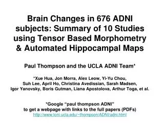
Brain Changes in 676 ADNI subjects: Summary of 10 Studiesusing Tensor Based Morphometry& Automated Hippocampal MapsPaul Thompson and the UCLA ADNI Team**Xue Hua, Jon Morra, Alex Leow, Yi-Yu Chou, Suh Lee, April Ho, Christina Avedissian, Sarah Madsen, Igor Yanovsky, Boris Gutman, Liana Apostolova, Arthur Toga, et al.*Google “paul thompson ADNI”to get a webpage with links to the full papers (PDFs)http://www.loni.ucla.edu/~thompson/ADNI/adni.html
Overview Mapped brain changes in 676 ADNI subjects • Tensor-based morphometry (gives 3D maps of rates of tissue loss) • Automated Hippocampal/Ventricular Mapping • 1000s of scans, no manual intervention Only need ~40 AD and 80 MCI subjects to detect 25% slowing of disease (10x better than best clinical score) Which MRI measures correlate best with clinical decline, and with CSF biomarkers (A-beta/Tau)? What is the best numeric summary of change from a 3D image? Is 3T better than 1.5T? How is power affected by pooling?
AD (N=104) versus Normal (N=157)- Mean Atrophy Rates - Hua, X. and Thompson, P.M. et al., 2009
MCI (N=254) versus Normal (N=157)- Mean Atrophy Rates - Hua, X. and Thompson, P.M. et al., 2009
AD (N=104) versus Normal (N=157)- Mean Atrophy Rates - Hua, X. and Thompson, P.M. et al., 2009
MCI (N=254) versus Normal (N=157)- Mean Atrophy Rates - Hua, X. and Thompson, P.M. et al., 2009
AD (N=104) versus Normal (N=157)- Mean Atrophy Rates - Hua, X. and Thompson, P.M. et al., 2009
MCI (N=254) versus Normal (N=157)- Mean Atrophy Rates - Hua, X. and Thompson, P.M. et al., 2009
MCI Converters Lose Tissue Faster Leow AD, Yanovsky I, Parikshak N, HuaX, LeeS, Toga AW, Jack CR, Bernstein MA, Britson PJ, Ward CP, Borowski B, Trojanowski JQ, Shaw L, Fleisher AS, Harvey D, Kornak J, Schuff N, Alexander GE, Weiner MW, Thompson PM (2009).Alzheimer’s Disease Neuroimaging Initiative: A One-year Follow up Study Correlating Degenerative Rates, Biomarkers and Cognition, NeuroImage, 2009 Apr 15;45(3):645-55.
Leow AD, Yanovsky I, Parikshak N, HuaX, LeeS, Toga AW, Jack CR, Bernstein MA, Britson PJ, Ward CP, Borowski B, Trojanowski JQ, Shaw L, Fleisher AS, Harvey D, Kornak J, Schuff N, Alexander GE, Weiner MW, Thompson PM (2009).Alzheimer’s Disease Neuroimaging Initiative: A One-year Follow up Study Correlating Degenerative Rates, Biomarkers and Cognition, NeuroImage, 2009 Apr 15;45(3):645-55.
Leow AD, Yanovsky I, Parikshak N, HuaX, LeeS, Toga AW, Jack CR, Bernstein MA, Britson PJ, Ward CP, Borowski B, Trojanowski JQ, Shaw L, Fleisher AS, Harvey D, Kornak J, Schuff N, Alexander GE, Weiner MW, Thompson PM (2009).Alzheimer’s Disease Neuroimaging Initiative: A One-year Follow up Study Correlating Degenerative Rates, Biomarkers and Cognition, NeuroImage, 2009 Apr 15;45(3):645-55.
For drug trials, want to summarize change in a Region-of-interest (ROI) Anatomical ROI based on temporal lobes Statistical ROI derived from an independent training sample of 22 AD patients Hua, X., Lee, S., Yanovsky, I., Leow, A.D., Chou, Y.Y., Ho, A.J., Gutman, B., Toga, A.W., Jack, C.R. Jr, Bernstein, M.A., Reiman, E.M., Harvey, D., Kornak, J., Schuff, N., Alexander, G.E., Fox, N.C., Weiner, M.W., Thompson, P.M. and the Alzheimer's Disease Neuroimaging Initiative, 2009. Optimizing Power to Track Brain Degeneration in Alzheimer's Disease and Mild Cognitive Impairment with Tensor-Based Morphometry: An ADNI Study of 515 Subjects. To be submitted to NeuroImage.
Statistical ROI reduces sample size by 15-50%; most helpful in MCI • Statistically-defined ROI outperforms the anatomically-defined temporal lobe ROI; extremely helpful in MCI, as it focuses on the part of the brain that is changing most* Hua, X., Lee, S., Yanovsky, I., Leow, A.D., Chou, Y.Y., Ho, A.J., Gutman, B., Toga, A.W., Jack, C.R. Jr, Bernstein, M.A., Reiman, E.M., Harvey, D., Kornak, J., Schuff, N., Alexander, G.E., Fox, N.C., Weiner, M.W., Thompson, P.M. and the Alzheimer's Disease Neuroimaging Initiative, 2009. Optimizing Power to Track Brain Degeneration in Alzheimer's Disease and Mild Cognitive Impairment with Tensor-Based Morphometry: An ADNI Study of 515 Subjects. To be submitted to NeuroImage.
Sample size estimates for a drug trial (= 48AD, 88 MCI)- does it matter what statistical threshold is used to define the region with greatest effect sizes for change? Hua, X. and Thompson, P.M. et al., 2009
Estimated sample sizes (n80)- needed to detect a 25% reduction in the mean annual change with a two-sided test and a=0.05 at 80% power, for a two-arm study • Sum-of-boxes Clinical Dementia Rating (CDR) gives best power among the clinical scores, but the TBM method is 10 times better Hua, X., Lee, S., Yanovsky, I., Leow, A.D., Chou, Y.Y., Ho, A.J., Gutman, B., Toga, A.W., Jack, C.R. Jr, Bernstein, M.A., Reiman, E.M., Harvey, D., Kornak, J., Schuff, N., Alexander, G.E., Fox, N.C., Weiner, M.W., Thompson, P.M. and the Alzheimer's Disease Neuroimaging Initiative, 2009. Optimizing Power to Track Brain Degeneration in Alzheimer's Disease and Mild Cognitive Impairment with Tensor-Based Morphometry: An ADNI Study of 515 Subjects. To be submitted to NeuroImage.
1.5T Is power better at 3T? 3 T
1.5T average 3 T average
More of the brain showed AD-accelerated tissue loss at 3T than at 1.5 T but with slightly weaker effect size (24 AD vs. 35 CTLs scanned at both field strengths) 1.5 T 3 T Ho, AJ., Hua, X., Lee, S., Leow, AD., Yanovsky, I., Gutman, B., Dinov, ID., Lepore, N., Stein, JL., Hojatkashani, C., Toga, AW., Jack, JR., Bernstein, MA., Reiman, EM., Harvey, D., Kornak, J., Schuff, N., Alexander, GE., Weiner, MW., Thompson, PM. Comparing 3 Tesla and 1.5 Tesla MRI for Tracking Alzheimer’s Disease Progression with Tensor-Based Morphometry.
Generated a statistical ROI for each field strength (slightly smaller at 3T) 1.5T ROI (darker gray) slightly larger than 3T ROI (lighter gray) Ho, AJ., Hua, X., Lee, S., Leow, AD., Yanovsky, I., Gutman, B., Dinov, ID., Lepore, N., Stein, JL., Hojatkashani, C., Toga, AW., Jack, JR., Bernstein, MA., Reiman, EM., Harvey, D., Kornak, J., Schuff, Norbert., Alexander, GE., Weiner, MW., Thompson, PM. Comparing 3 Tesla and 1.5 Tesla MRI for Tracking Alzheimer’s Disease Progression with Tensor-Based Morphometry.
MCI: Power slightly worse at 3T, similar in AD N80 = Minimal Sample Sizes, per diagnostic group, to detect 25% slowing of the mean atrophic rate (with 80% power, alpha = 0.05). Ho, AJ., Hua, X., Lee, S., Leow, AD., Yanovsky, I., Gutman, B., Dinov, ID., Lepore, N., Stein, JL., Hojatkashani, C., Toga, AW., Jack, JR., Bernstein, MA., Reiman, EM., Harvey, D., Kornak, J., Schuff, Norbert., Alexander, GE., Weiner, MW., Thompson, PM. Comparing 3 Tesla and 1.5 Tesla MRI for Tracking Alzheimer’s Disease Progression with Tensor-Based Morphometry.
Mixing 3T and 1.5T scanners - Power did not degrade at all when 25% of the scanners were 3T 3T ONLY 159 1.5T ONLY 134 134 103 107 48 58 52 37 37 MIX IS OK Ho, AJ., Hua, X., Lee, S., Leow, AD., Yanovsky, I., Gutman, B., Dinov, ID., Lepore, N., Stein, JL., Hojatkashani, C., Toga, AW., Jack, JR., Bernstein, MA., Reiman, EM., Harvey, D., Kornak, J., Schuff, Norbert., Alexander, GE., Weiner, MW., Thompson, PM. Comparing 3 Tesla and 1.5 Tesla MRI for Tracking Alzheimer’s Disease Progression with Tensor-Based Morphometry.
Morra JH, Tu Z, Apostolova LG, Green A, Toga AW, Thompson PM (2009). Comparison of Adaboost and Support Vector Machines for Detecting Alzheimer’s Disease through Automated Hippocampal Segmentation, IEEE Transactions on Medical Imaging, in press.
Morra JH, Tu Z, Apostolova LG, Green A, Toga AW, Thompson PM (2009). Comparison of Adaboost and Support Vector Machines for Detecting Alzheimer’s Disease through Automated Hippocampal Segmentation, IEEE Transactions on Medical Imaging, in press.
HP Loss Rates (980 scans) Morra, J., Tu, Z., Apostolova, L.G., Green, A., Avedissian, C., Madsen, S., Parikshak, N., Hua, X., Toga, A., Jack, C., Schuff, N., Weiner, M., Thompson, P., 2008. Automated Mapping of Hippocampal Atrophy in 1-Year Repeat MRI Data in 490 Subjects with Alzheimer’s Disease, Mild Cognitive Impairment, and Elderly Controls. Neuroimage.
ApoE4+ atrophied 2-3% faster Morra, J., Tu, Z., Apostolova, L.G., Green, A., Avedissian, C., Madsen, S., Parikshak, N., Hua, X., Toga, A., Jack, C., Schuff, N., Weiner, M., Thompson, P., 2008. Automated Mapping of Hippocampal Atrophy in 1-Year Repeat MRI Data in 490 Subjects with Alzheimer’s Disease, Mild Cognitive Impairment, and Elderly Controls. Neuroimage.
AT BASELINE, GROUP DIFFERENCES ARE MAPPED: Morra, J., Tu, Z., Apostolova, L.G., Green, A., Avedissian, C., Madsen, S., Parikshak, N., Hua, X., Toga, A., Jack, C., Schuff, N., Weiner, M., Thompson, P., 2008. Automated Mapping of Hippocampal Atrophy in 1-Year Repeat MRI Data in 490 Subjects with Alzheimer’s Disease, Mild Cognitive Impairment, and Elderly Controls. Neuroimage.
Significance Maps BASELINE ATROPHY CORRELATES WITH:
Chou YY, Leporé N, Avedissian C, Madsen S, Parikshak N, Hua X, Trojanowski J, Shaw L, Weiner M, Toga A, Thompson PM (2009). Mapping Correlations between Ventricular Expansion, and CSF Amyloid & Tau Biomarkers in 240 Subjects with Alzheimer’s Disease, Mild Cognitive Impairment & Elderly Controls, NeuroImage. 2009 Feb 21.
Chou YY, Leporé N, Avedissian C, Madsen SK, Parikshak N, Hua X, Trojanowski JQ, Shaw L, Weiner MW, Toga AW, Thompson PM (2009). Mapping Correlations between Ventricular Expansion, and CSF Amyloid & Tau Biomarkers in 240 Subjects with Alzheimer’s Disease, Mild Cognitive Impairment and Elderly Controls, NeuroImage. 2009 Feb 21. [Epub ahead of print]
Summary All MRI measures correlate well with CSF biomarkers, clinical decline, and predict future conversion to AD. TBM needs 50 AD and 75 MCI subjects to detect 25% slowing of disease (10x better than best clinical score) All maps show focal effects - interesting that these statistically guided ROIs will give much better numeric summaries of change (15-50% reductions in sample size) Mixing 1.5T and 3T scanners is not a problem; but 3T was slightly worse for tracking change in MCI