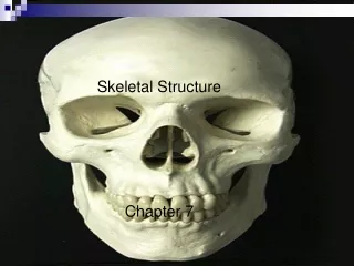
Chapter 7: Skeletal Tissues
550 likes | 574 Views
Learn the functions of bones, types of bone tissues, and microscopic structure of bone. Discover how bones support, protect, and facilitate movement in the body.

Chapter 7: Skeletal Tissues
E N D
Presentation Transcript
FUNCTIONS OF BONE Support: bones form the framework of the body and contribute to the shape, alignment, and positioning of body parts; ligaments help hold bones together (Figure 7-1) Protection: bony “boxes” protect the delicate structures they enclose Movement: bones and their joints constitute levers that move as muscles contract Mineral storage: bones are the major reservoir for calcium, phosphorus, and other minerals Hematopoiesis: blood cell formation is carried out by myeloid tissue
TYPES OF BONES Five major types of structural bones (Figure 7-2) Long bones Short bones Flat bones Irregular bones Sesamoid bones Bones serve various needs, and their size, shape, and appearance vary to meet those needs Bones vary in the proportion of compact and cancellous (spongy) bone; compact bone is dense and solid in appearance, whereas cancellous bone is characterized by open space partially filled with needlelike structures
TYPES OF BONES (cont.) Parts of a long bone (Figure 7-3) Diaphysis Main shaft of a long bone Hollow, cylindrical shape and thick compact bone Function is to provide strong support without cumbersome weight Epiphyses Both ends of a long bone; made of cancellous bone filled with marrow Bulbous shape Function is to provide attachments for muscles and give stability to joints
TYPES OF BONES (cont.) Articular cartilage Layer of hyaline cartilage that covers the articular surface of epiphyses Function is to cushion jolts and blows Periosteum Dense, white fibrous membrane that covers bone Attaches tendons firmly to bones Contains cells that form and destroy bone Contains blood vessels important in growth and repair Contains blood vessels that send branches into bone Essential for bone cell survival and bone formation
TYPES OF BONES (cont.) Medullary (or marrow) cavity Tubelike, hollow space in the diaphysis Filled with yellow marrow in adults Endosteum: thin, fibrous membrane that lines the medullary cavity
TYPES OF BONES (cont.) Parts of a flat bone Inner portion is cancellous bone covered on the outside with compact bone Cranial flat bones have an internal and external table of compact bone and an inner cancellous region called the diploë (Figure 7-4) Bones are covered with periosteum and lined with endosteum, such as in a long bone Other flat bones, short bones, and irregular bones have features similar to the cranial bones Spaces inside the cancellous bone of short, flat, irregular and sesamoid bones are filled with red marrow
BONE TISSUE Most distinctive form of connective tissue Extracellular components are hard and calcified Rigidity of bone gives it supportive and protective functions Tensile strength nearly equal to that of cast iron at less than one third the weight
BONE TISSUE (cont.) Composition of bone matrix Inorganic salts Hydroxyapatite: crystals of calcium and phosphate contribute to bone hardness Magnesium, sodium, sulfate, and fluoride are also found in bone Organic matrix Composite of collagenous fibers and an amorphous mixture of protein and polysaccharides called ground substance Chondroitin sulfate (compression) and glucosamine (growth and repair) Adds to overall strength of bone and gives some degree of resilience to bone
MICROSCOPIC STRUCTURE OF BONE Compact bone (Figure 7-5) – 80% Contains many cylinder-shaped structural units called osteons, or haversian systems (Figure 7-6) Osteons surround central (osteonal or haversian) canals that run lengthwise through bone and are connected by transverse (Volkmann) canals Living bone cells are located in these units, which constitute the structural framework of compact bone Osteons permit delivery of nutrients and removal of waste products
MICROSCOPIC STRUCTURE OF BONE (cont.) Structures that make up each osteon Lamellae Concentric: cylinder-shaped layers of calcified matrix around the central canal Interstitial: layers of bone matrix between the osteons; leftover from previous osteons Circumferential: few layers of bone matrix that surround all the osteons; run along the outer circumference of a bone and inner circumference (boundary of medullary cavity) of a bone
MICROSCOPIC STRUCTURE OF BONE (cont.) Structures that make up each osteon (cont.) Lacunae: small spaces containing tissue fluid in which bone cells are located between hard layers of the lamella Canaliculi: ultra-small canals radiating in all directions from the lacunae and connecting them to each other and to the central canal Central (osteonal or Haversian) canal: extends lengthwise through the center of each osteon; contains blood vessels and lymphatic vessels
MICROSCOPIC STRUCTURE OF BONE (cont.) Cancellous bone (Figure 7-6) – 20% No osteons in cancellous bone; it has trabeculae instead Nutrients are delivered and waste products removed by diffusion through tiny canaliculi Bony branches (trabeculae) are arranged along lines of stress to enhance the bone’s strength (Figure 7-7) Blood supply Bone cells are metabolically active and need a blood supply, which comes from the bone marrow in the internal medullary cavity of cancellous bone
MICROSCOPIC STRUCTURE OF BONE (cont.) Types of bone cells Osteoblasts (Figure 7-8) Bone-forming cells found in all bone surfaces Small cells synthesize and secrete osteoid, an important part of the ground substance
MICROSCOPIC STRUCTURE OF BONE (cont.) Types of bone cells Osteoclasts Giant multinucleated cells Responsible for the active erosion of bone minerals Contain large numbers of mitochondria and lysosomes Osteocytes: mature, nondividing osteoblasts surrounded by matrix and lying within lacunae (Figure 7-9)
BONE MARROW Type of soft, diffuse connective tissue; called myeloid tissue Site for the production of blood cells Found in the medullary cavities of long bones and in the spaces of spongy bone
BONE MARROW (cont.) Two types of marrow occur during a person’s lifetime Red marrow Found in virtually all bones in an infant’s or child’s body Produces red blood cells Yellow marrow As an individual ages, red marrow is replaced by yellow marrow Marrow cells become saturated with fat and are no longer active in blood cell production
BONE MARROW (cont.) The main bones in an adult that still contain red marrow include the ribs, bodies of the vertebrae, humerus, pelvis, and femur Yellow marrow can change to red marrow during times of decreased blood supply, such as anemia, exposure to radiation, and certain diseases
REGULATION OF BLOOD CALCIUM LEVELS Skeletal system is a storehouse for about 98% of body calcium reserves Helps maintain constancy of blood calcium levels Calcium is mobilized and moves in and out of blood during bone remodeling During bone formation, osteoblasts remove calcium from blood and lower circulating levels During breakdown of bone, osteoclasts release calcium into blood and increase circulating levels
REGULATION OF BLOOD CALCIUM LEVELS (cont.) Homeostasis of calcium ion concentration essential for the following: Bone formation, remodeling, and repair Blood clotting Transmission of nerve impulses Maintenance of skeletal and cardiac muscle contraction pH regulation
REGULATION OF BLOOD CALCIUM LEVELS (cont.) Mechanisms of calcium homeostasis (Figure 7-10) Parathyroid hormone Primary regulator of calcium homeostasis Stimulates osteoclasts to initiate breakdown of bone matrix and increase blood calcium levels Increases renal absorption of calcium from urine Stimulates vitamin D synthesis Increases Blood [Ca++] levels When blood passing through the parathyroid gland is sufficient, PTH secretion is stopped
REGULATION OF BLOOD CALCIUM LEVELS (cont.) Mechanisms of calcium homeostasis Calcitonin Protein hormone produced in the thyroid gland Produced in response to high blood calcium levels Stimulates bone deposition by osteoblasts Inhibits osteoclast activity Far less important in homeostasis of blood calcium levels than is parathyroid hormone Decreases Blood [Ca++]
DEVELOPMENT OF BONES Osteogenesis: development of bone from small cartilage model to adult bone (Figure 7-11) Intramembranous ossification Occurs within a connective tissue membrane Flat bones begin when groups of cells differentiate into osteoblasts Osteoblasts are clustered together in ossification center Osteoblasts secrete matrix material and collagenous fibrils
DEVELOPMENT OF BONES (cont.) Intramembranous ossification Large amounts of ground substance accumulate around each osteoblast Collagenous fibers become embedded in the ground substance and constitute the bone matrix Bone matrix calcifies when calcium salts are deposited Trabeculae appear and join in a network to form spongy bone Appositional growth occurs by adding osseous tissue
DEVELOPMENT OF BONES (cont.) Endochondral ossification (Figure 7-12) Most bones begin as a cartilage model with bone formation spreading essentially from the center to the ends Periosteum develops and enlarges to produce a collar of bone Primary ossification center forms (Figure 7-13) Blood vessel enters the cartilage model at the midpoint of the diaphysis Bone grows in length as endochondral ossification progresses from the diaphysis toward each epiphysis (Figure 7-14) Secondary ossification centers appear in the epiphysis, and bone growth proceeds toward the diaphysis Epiphyseal plate remains between the diaphysis and each epiphysis until bone growth in length is complete
DEVELOPMENT OF BONES (cont.) Epiphyseal plate is composed of four layers (Figures 7-15 and 7-16) “Resting” cartilage cells: point of attachment joining the epiphysis to the shaft Zone of proliferation: cartilage cells undergoing active mitosis, which causes the layer to thicken and the plate to increase in length Zone of hypertrophy: older, enlarged cells undergoing degenerative changes associated with calcium deposition Zone of calcification: dead or dying cartilage cells undergoing rapid calcification
DEVELOPMENT OF BONES (cont.) Epiphyseal plate can be a site for bone fractures in young people (Figure 7-17) Long bones grow in both length (interstitial growth) and diameter (appositional growth) (Figure 7-18) Why care about an epiphyseal plate fracture?
BONE REMODELING Primary osteons develop within early woven bone (Figure 7-19) Conelike or tubelike space is hollowed out by osteoclasts Osteoblasts in the endosteum that lines the tube begin forming layers (lamellae) that trap osteocytes between layers A central canal is left for the blood and lymphatic vessels and nerves Bones grow in length and diameter by the combined action of osteoclasts and osteoblasts Osteoclasts enlarge the diameter of the medullary cavity Osteoblasts from the periosteum build new bone around the outside of the bone Between 35-40 bone loss surpasses bone growth Mechanical stress, such as physical activity, strengthens bone
REPAIR OF BONE FRACTURES Fracture: break in the continuity of a bone Fracture healing (Figure 7-20) Fracture tears and destroys blood vessels that carry nutrients to osteocytes Vascular damage initiates repair sequence Fracture hematoma: blood clot occurring immediately after the fracture, which is then resorbed and replaced by callus Callus: special repair tissue that stabilizes the bone so healing can occur and bone replaces callus
CARTILAGE Characteristics Avascular connective tissue Fibers of cartilage are embedded in a firm gel Has the flexibility of firm plastic No canal system or blood vessels Chondrocytes receive oxygen and nutrients by diffusion Perichondrium: fibrous covering of the cartilage Cartilage types differ because of the amount of matrix present and the amounts of elastic and collagenous fibers
CARTILAGE (cont.) Types of cartilage (Figure 7-21) Hyaline cartilage Most common type Covers the articular surfaces of bones Forms the costal cartilages, cartilage rings in the trachea, bronchi of the lungs, and the tip of the nose Forms from special cells in chondrification centers, which secrete matrix material Chondrocytes are isolated into lacunae