Download
1 / 62
620 likes | 650 Views
Learn about femoral artery identification, groin hemostasis methods, patient care post-procedure, complications, PVD, risk factors, indications for angiography, and more.

E N D
GROIN MANAGEMENTby Josh Marshall, RTR, RCIS.Trish O’Sullivan, RN. • OBJECTIVES • IDENTIFY FEMORAL ARTERY, AND WHY IT’S COMMONLY USED FOR ACCESS. LOOK AT OTHER ACCESS SITES. • CURRENT METHODS FOR GROIN HEMOSTASIS • MANAGEMENT OF PATIENT FOLLOWING SHEATH PLACEMENT AND REMOVAL • COMPLICATIONS AND WARNING SIGNS • POST PROCEDURE CARE FROM A NURSING PERSPECTIVE
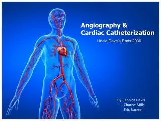
What is Angiography? • An x-ray exam of the blood vessels using contrast media to diagnose blockages and other blood vessel problems
What is PVD • PVD is a disease of the blood vessels outside of the heart and brain. It is a common circulation problem in which arteries that carry blood to the legs or arms become narrowed or clogged. PVD is also referred to as peripheral arterial disease or PAD. • Atherosclerosis is the predominant factor contributing to PAD. It is gradual, progressive and irreversible process directly related to risk factors.
PVD Uncontrollable risk factors • Diabetes • Age • Gender • Family history
PVD Risk factors…Cont • Modifiable risk factors • Cigarette smoking • High lipid diet • HTN • Sedentary lifestyle
Signs and Symptoms • Pain • Pallor • Pulselessness • Coolness • Paresethesia • Paralysis • Pallor
Objective signs in patients with PVD • Leg hair loss • Callus formation • Ulceration • Tissue loss • Gangrene • Impotence
Indications for Angiography • Diagnosis of PVD • Pre op definition of vascular anatomy. • Access for endovascular therapy which include: • PTA • STENTING • ATHRECTOMY • THROMBOLYTIC THERAPY • EMBOLIZATION • (These procedures are often done in conjunction with one another)
Athrectomy • CSI Diamond back is used to change vessel compliance and allow optimal results from PTA balloons, by de-bulking hard clacific and other types of blockages from blood vessels
Pre procedure Management • Indication for study • Review Med Surg history • Lab data • Current meds • Allergies • NPO status • ABI • Consent • Education
On the with the case!!! • Pt prepped and draped. • attached to Monitor system. • Local anesthetic for access site. • Medications given for sedation or necessary pre-treatment.
Femoral access • Most common method is the Seldinger technique. Used mostly in the cath lab • 1-2cm below the inguinal ligament in the common femoral above the bifurcation of profunda and SFA • Cutdown technique…. not so common
Why we use the femoral artery • Large caliber vessel anywhere from 4-6mm large enough to use large sheaths. • Compressible over the femoral head • high sticks can cause retroperitoneal bleeds and low sticks have been linked to psuedoaneurysm, and AV fistulas • Body habitus factors into sheath placement at times
Other access sites….. • Brachial • Radial
Catheter placement For an Ao gram is just above The Renal arteries.
Antegrade V.S. Retrograde • Retrograde: going against the flow of blood. ( as seen with heart caths and most vasculars) • Antegrade: Going with the flow of blood. (as seen with vascular cases) Note that an arterial stick above the inguinal ligament could result in a life threatening retroperitoneal bleed. • Pay close attention to patients with antegrade sticks!!!! Question back pain and Changes in BP/vital signs.
Reasons for complications • Technique (ie. multiple sticks, Lack of pulse assessment) • Antiplatlet & Anti-thombotic adjunctive treatments (Important to note patient INR results) • Patient complexity • Female gender • Obesity vs. low Body mass index • Age • Catheter size • Bad luck
Facts • Estimated that 5-10% of all arterial access will result in a hematoma, infection, pseudoaneurysm, or retroperitoneal bleed. • Most complications are from initial puncture or insertion of sheath • Specific problems include…vessel thrombosis, dissection, poorly controlled bleeding at site • Management of these problems is challenging with the use of anticoagulation
How to minimize risk!!! • Patient history- past procedures (scar tissue) surgeries, and response to medications, INR results 1.5 and below is considered safe for access. • Knowledge of co morbidities such as: • diabetes • Hypertension • Stroke • Smoking • PVD • Elderly, females, and low body weight
Co-morbid conditions • Diabetes- patients usually have small diffuse arteries which are prone to disease and are also at risk for infection • Hypertension-Are at risk for bleeding with sheath insertion and removal. Anti-hypertension medication may be required to stabilize the blood pressure prior to sheath removal. • Multiple procedures- Re-access is not only uncomfortable but difficult for operator to penetrate the subcutaneous scar tissue, which could cause the sheath to bend or break upon insertion into artery
Co-morbid Conditions Cont. • Thin patients- minimal tissue for secure sheath placement and homeostasis post sheath removal can be difficult • Atherosclerosis- patients with extensive vascular disease have brittle or friable vessels, which are not only difficult to access but can also lead to distal embolization due to calcification. • Tortuous iliac artery- requires careful sheath insertion and a longer sheath may be necessary to bypass the the tortuosity. • Obesity-the position of the inguinal ligament to skin crease, may be misleading. Often large panis must be taped up or moved for access, and groin hold can be challenging for operator • Known PVD, or CAD. This information will help make assessment of access sites easier to understand..
Distal Aortic disease Femoral pulses could be weak or absent, ultrasound may be used for access
Methods of hemostasis • Manual compression (gold standard) • Mechanical compression (femstop) • Vascular closure devices such as Angioseal, Mynx • Hemostatic patches (D-stat dry patches)
Rules for manual compression • check the site for hematoma and distal pulses. Post Tib and DP for groin access, and radial for brachial access. • Manual compression above sheath access point. 15-20 min is normally adequate. • Note patients starting BP and heart rate. Patients can vagal during sheath pulls. ( atropine , fluids, trendelenburg) • If hematoma occurs firm compression is best first response.
Distal Pulse Check Sites • Dorsal Pedis • Post Tib • Pop • Radial
For antegrade sticks hold above And on the site
Use of Vascular closure devices in the Lab. • Reduce time to ambulate • Reduce time to discharge • Improve Patient satisfaction • Can Reduce cost
Pre angioseal Femoral angiogram xxxxxxxxxxxxxx xxxxxxxxxx xxxxxxxxxxx xxxxxxxxxxxxx Placement depends upon: 1. Sheath in non-diseased segment of femoral artery 2. Above the bifurcation of the SFA, and Profunda, 3. Vessel is of large enough caliber.
Vascular closure device limitations • Pvd • Bifurcation sticks • Re-access issues • Small femoral vessel • Obesity/ low BMI • Double wall sticks
Femoral artery complications with vascular closure device • Bleeding • Embolization: device or collagen • Direct vessel trauma • Vessel occlusion • infection
Predictors of access site complications • Anticoagulant used- heparin, GP11b/111a, high INR’s • Patient demographics/ underlying co-morbidities • Sheath insertion technique, multiple sheath insertions. • Sheath removal technique • Poor manual compression • Improper use of mechanical devices • Closure device failure
Angiographic complications • Neurological • Puncture site ( more detail to follow) • Catheter thrombosis • Contrast induced allergy/ anaphylaxis • Contrast induced renal failure • infection
Hematoma • Swelling, enlarged area usually around the access site, may or may not be associated with strong bleeding • Possible source: • Multiple sticks, posterior wall stick • Inadequate manual compression • Action • Firm compression over site, or possible surgical intervention
Retroperitoneal bleed • Although rare 0.75%, is a potentially fatal complication • The bleeding occurs inside the peritoneal cavity. Patients often complain of back/ flank pain and will have changes in vital sings ie.. Drop in Bp, and changes in mentation: confusion, lethargy • Sources: high stick, posterior wall stick, female gender, low body surface area