Download
1 / 10
0 likes | 20 Views
Lower-Limb-Anatomy-An-Exploration Karthick. S, DT.

E N D
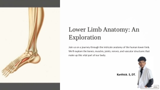
Lower Limb Anatomy: An Exploration Join us on a journey through the intricate anatomy of the human lower limb. We'll explore the bones, muscles, joints, nerves, and vascular structures that make up this vital part of our body. Karthick. S, DT.
The Bones of the Lower Limb Femur Tibia and Fibula Tarsals, Metatarsals, and Phalanges The longest and strongest bone in the body. It forms the thigh and connects to the hip and knee joints. Two bones that form the lower leg, with the tibia bearing the majority of weight. They connect to the knee and ankle joints. The bones that make up the foot, providing stability and support for walking and balance.
Muscles of the Lower Extremity Quadriceps Hamstrings The large muscle group on the front of the thigh, responsible for extending the knee. Three muscles located on the back of the thigh, responsible for flexing the knee and extending the hip. Gastrocnemius and Soleus Tibialis Anterior A muscle located on the front of the shin, responsible for dorsiflexing the foot and inverting the ankle. The calf muscles, responsible for plantarflexing the foot and propelling us forward during walking.
Joints and Ligaments of the Lower Limb Hip Joint Knee Joint A ball-and-socket joint that allows for a wide range of motion, connecting the femur to the pelvis. A hinge joint that allows for flexion and extension of the leg, connecting the femur to the tibia and fibula. Ankle Joint A hinge joint that allows for dorsiflexion and plantarflexion of the foot, connecting the tibia and fibula to the talus bone.
Nerves Innervating the Lower Limb Femoral Nerve 1 Supplies the anterior thigh muscles and skin of the anterior thigh and medial leg. Sciatic Nerve 2 The largest nerve in the body, branching into the tibial and common peroneal nerves, supplying the posterior thigh, leg, and foot. Tibial Nerve 3 Supplies the posterior leg and plantar muscles of the foot. Common Peroneal Nerve 4 Supplies the anterior and lateral leg and foot muscles, responsible for dorsiflexion and eversion.
Vascular Supply to the Lower Extremity Femoral Artery Popliteal Artery Anterior Tibial Artery Posterior Tibial Artery The main artery supplying the lower limb, branching into the popliteal artery, anterior tibial artery, and posterior tibial artery. Supplies the knee and calf muscles, branching into the anterior and posterior tibial arteries. Supplies the anterior leg and foot muscles. Supplies the posterior leg and plantar muscles of the foot.
The Arches of the Foot and Gait Mechanics Medial Longitudinal Arch 1 Provides support and shock absorption during walking. Lateral Longitudinal Arch 2 Supports the outer side of the foot and helps with balance. Transverse Arch 3 Supports the middle of the foot and distributes weight evenly.
Common Injuries and Conditions of the Lower Limb 1 2 ACL Tear Ankle Sprain A common knee injury, often caused by a sudden twisting or hyperextension movement. An injury to the ligaments that support the ankle joint, often caused by a sudden twist or roll. 3 4 Achilles Tendinitis Plantar Fasciitis Inflammation of the Achilles tendon, which connects the calf muscles to the heel bone. Inflammation of the plantar fascia, a thick band of tissue on the bottom of the foot.
Diagnostic Techniques for Lower Limb Evaluation Physical Examination 1 A comprehensive assessment of the patient's symptoms, range of motion, and gait. Imaging Studies 2 X-rays, MRIs, and ultrasounds to visualize the bones, muscles, ligaments, and tendons. Electromyography (EMG) 3 A test that measures the electrical activity of muscles, used to diagnose nerve damage.