Download

1 / 28
280 likes | 412 Views
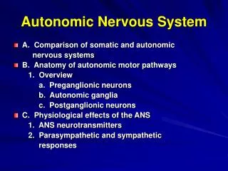
12. The Central Nervous System: Part A. Regions and Organization of the CNS. Adult brain regions Cerebral hemispheres Diencephalon Brain stem (midbrain, pons, and medulla) Cerebellum. Cerebral hemisphere. Diencephalon. Cerebellum. Brain stem. • Midbrain. • Pons. • Medulla oblongata.

E N D
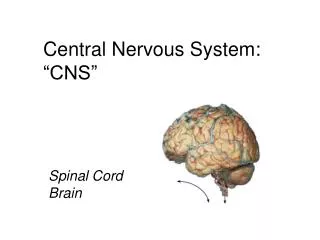
12 The Central Nervous System: Part A
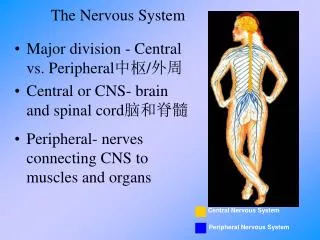
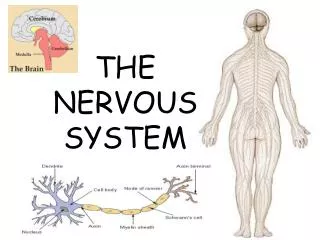
Regions and Organization of the CNS • Adult brain regions • Cerebral hemispheres • Diencephalon • Brain stem (midbrain, pons, and medulla) • Cerebellum
Cerebral hemisphere Diencephalon Cerebellum Brain stem • Midbrain • Pons • Medullaoblongata (d) Birth Figure 12.3d
Left cerebral hemisphere Transverse cerebral fissure Brain stem Cerebellum (d) Figure 12.6d
Lateral ventricle Septum pellucidum Anterior horn Posterior horn Inferior horn Interventricular foramen Lateral aperture Median aperture Third ventricle Inferior horn Lateral aperture Cerebral aqueduct Fourth ventricle Central canal (a) Anterior view (b) Left lateral view Figure 12.5
Precentral gyrus Central sulcus Postcentral gyrus Frontal lobe Parietal lobe Parieto-occipital sulcus (on medial surface of hemisphere) Lateral sulcus Occipital lobe Temporal lobe Transverse cerebral fissure Cerebellum Pons Medulla oblongata Fissure Spinal cord (a deep sulcus) Gyrus Cortex (gray matter) Sulcus White matter (a) Figure 12.6a
Anterior Longitudinal fissure Frontal lobe Cerebral veins and arteries covered by arachnoid mater Parietal lobe Right cerebral hemisphere Left cerebral hemisphere Occipital lobe Posterior (c) Figure 12.6c
Anterior Cerebral cortex Cerebral white matter Corpus callosum Anterior horn of lateral ventricle Caudate nucleus Putamen Lentiform nucleus Globus pallidus Thalamus Tail of caudate nucleus Third ventricle Inferior horn of lateral ventricle (b) Posterior Figure 12.11b (1 of 2)
Commissural fibers (corpus callosum) Longitudinal fissure Superior Lateral ventricle Association fibers Basal nuclei • Caudate Corona radiata • Putamen • Globuspallidus Fornix Internal capsule Thalamus Gray matter Third ventricle White matter Projection fibers Pons Decussation of pyramids Medulla oblongata (a) Figure 12.10a
Frontal lobe Olfactory bulb (synapse point of cranial nerve I) Optic chiasma Optic nerve (II) Optic tract Mammillary body Midbrain Pons Temporal lobe Medulla oblongata Cerebellum Spinal cord Figure 12.14
Motor areas Sensory areas and related association areas Central sulcus Primary motor cortex Primary somatosensory cortex Premotor cortex Somatic sensation Frontal eye field Somatosensory association cortex Broca’s area (outlined by dashes) Gustatory cortex (in insula) Taste Prefrontal cortex Wernicke’s area (outlined by dashes) Working memory for spatial tasks Executive area for task management Primary visual cortex Working memory for object-recall tasks Vision Visual association area Solving complex, multitask problems Auditory association area Hearing Primary auditory cortex (a) Lateral view, left cerebral hemisphere Motor association cortex Primary sensory cortex Primary motor cortex Sensory association cortex Multimodal association cortex Figure 12.8a
Posterior Motor Anterior Motor map in precentral gyrus Toes Jaw Primary motor cortex (precentral gyrus) Tongue Swallowing Figure 12.9
Posterior Sensory Anterior Sensory map in postcentral gyrus Genitals Primary somato- sensory cortex (postcentral gyrus) Intra- abdominal Figure 12.9
Commissural fibers (corpus callosum) Longitudinal fissure Superior Lateral ventricle Association fibers Basal nuclei • Caudate Corona radiata • Putamen • Globuspallidus Fornix Internal capsule Thalamus Gray matter Third ventricle White matter Projection fibers Pons Decussation of pyramids Medulla oblongata (a) Figure 12.10a
Basal Nuclei (Ganglia) • Subcortical nuclei • Consists of the corpus striatum • Caudate nucleus • Lentiform nucleus (putamen + globus pallidus) • Functionally associated with the subthalamic nuclei (diencephalon) and the substantia nigra (midbrain)
Fibers of corona radiata Caudate nucleus Thalamus Lentiform nucleus • Putamen • Globus pallidus (deep to putamen) Tail of caudate nucleus Corpus striatum Projection fibers run deep to lentiform nucleus (a) Figure 12.11a
Anterior Cerebral cortex Cerebral white matter Corpus callosum Anterior horn of lateral ventricle Caudate nucleus Putamen Lentiform nucleus Globus pallidus Thalamus Tail of caudate nucleus Third ventricle Inferior horn of lateral ventricle (b) Posterior Figure 12.11b (1 of 2)
Cerebral cortex Cerebral white matter Corpus callosum Anterior horn of lateral ventricle Caudate nucleus Lentiform nucleus Thalamus Third ventricle Inferior horn of lateral ventricle (b) Figure 12.11b (2 of 2)
Cerebral hemisphere Diencephalon Cerebellum Brain stem • Midbrain • Pons • Medullaoblongata (d) Birth Figure 12.3d
Cerebral hemisphere Septum pellucidum Corpus callosum Interthalamic adhesion (intermediate mass of thalamus) Fornix Choroid plexus Thalamus (encloses third ventricle) Interven- tricular foramen Posterior commissure Pineal gland (part of epithalamus) Anterior commissure Corpora quadrigemina Mid- brain Cerebral aqueduct Hypothalamus Optic chiasma Arbor vitae (of cerebellum) Pituitary gland Fourth ventricle Mammillary body Choroid plexus Pons Cerebellum Medulla oblongata Spinal cord Figure 12.12
Frontal lobe Olfactory bulb (synapse point of cranial nerve I) Optic chiasma Optic nerve (II) Optic tract Mammillary body Midbrain Pons Temporal lobe Medulla oblongata Cerebellum Spinal cord Figure 12.14
Fourth ventricle Solitary nucleus Choroid plexus Hypoglossal nucleus (XII) Dorsal motor nucleus of vagus (X) Vestibular nuclear complex (VIII) Inferior cerebellar peduncle Cochlear nuclei (VIII) Lateral nuclear group Nucleus ambiguus Medial nuclear group Reticular formation Inferior olivary nucleus Raphe nucleus Pyramid Medial lemniscus (c) Medulla oblongata Figure 12.16c
Anterior lobe Cerebellar cortex Arbor vitae Cerebellar peduncles Posterior lobe • Superior • Middle Choroid plexus of fourth ventricle • Inferior Medulla oblongata Flocculonodular lobe (b) Figure 12.17b
Fiber tracts connecting limbic system structures Septum pellucidum Diencephalic structures of the limbic system Corpus callosum •Fornix •Anterior thalamic nuclei (flanking 3rd ventricle) •Anterior commissure Cerebral struc- tures of the limbic system •Hypothalamus •Mammillary body •Cingulate gyrus •Septal nuclei •Amygdala •Hippocampus •Dentate gyrus •Parahippocampal gyrus Olfactory bulb Figure 12.18
Lateral ventricle Septum pellucidum Anterior horn Posterior horn Inferior horn Interventricular foramen Lateral aperture Median aperture Third ventricle Inferior horn Lateral aperture Cerebral aqueduct Fourth ventricle Central canal (a) Anterior view (b) Left lateral view Figure 12.5
Superior sagittal sinus 4 Choroid plexus Arachnoid villus Interventricular foramen Subarachnoid space Arachnoid mater Meningeal dura mater Periosteal dura mater 1 Right lateral ventricle (deep to cut) Choroid plexus of fourth ventricle 3 Third ventricle 1 CSF is produced by the choroid plexus of each ventricle. Cerebral aqueduct Lateral aperture 2 CSF flows through the ventricles and into the subarachnoid space via the median and lateral apertures. Some CSF flows through the central canal of the spinal cord. Fourth ventricle Median aperture 2 Central canal of spinal cord 3 CSF flows through the subarachnoid space. (a) CSF circulation 4 CSF is absorbed into the dural venous sinuses via the arachnoid villi. Figure 12.26a
Ependymal cells Capillary Section of choroid plexus Connective tissue of pia mater Wastes and unnecessary solutes absorbed CSF forms as a filtrate containing glucose, oxygen, vitamins, and ions (Na+, Cl–, Mg2+, etc.) Cavity of ventricle (b) CSF formation by choroid plexuses Figure 12.26b
Skin of scalp Periosteum Bone of skull Dura mater Periosteal Meningeal Superior sagittal sinus Arachnoid mater Pia mater Arachnoid villus Subdural space Blood vessel Falx cerebri (in longitudinal fissure only) Subarachnoid space Figure 12.24