Download
1 / 8
80 likes | 299 Views
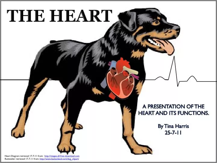
THE HEART. A PRESENTATION OF THE HEART AND ITS FUNCTIONS. By Tina Harris 25 -7-11. Heart Diagram retrieved 17-7-11 from: http://images.all-free-download.com Rottweiler retrieved 17-7-11 from: http://www.buttonland.com/dog_clipart/. The Heart.

E N D
THE HEART A PRESENTATION OF THE HEART AND ITS FUNCTIONS. By Tina Harris 25-7-11 Heart Diagram retrieved 17-7-11 from: http://images.all-free-download.com Rottweiler retrieved 17-7-11 from: http://www.buttonland.com/dog_clipart/
TheHeart • The Heart is a hollow muscular organ that lies within in the thoracic cavity. • The heart consists of two pumps that operate simultaneously to pump blood into the Pulmonary (Lungs) and Systemic (Body) circulatory systems. • The Heart is separated into left and right sides. • Each side has two chambers, the Atrium and Ventricle. Heart Diagram retrieved 17-7-11 from : http://www.zimbio.com/Emo/articles/KjErLtukm_K/heart+diagram+enchanted+learning
CranialVenaCava (From Body) • Arteries takebloodawayfromthe heartand Veins carrybloodbackto the heart. • Deoxygenated blood from the body enters the Right Atrium via the Vena Cava, passes through the Right Atrioventricular valve. • The blood exits theheartviathe Pulmonary Artery,andistakentotheLungs (pulmonary), where itis oxygenated viagasexchange. Pulmonary Artery (To Lungs) RightAtrioventricularvalve CaudalVenaCava (From Body) Heart Diagram retrieved 17-7-11 from : http://www.zimbio.com/Emo/articles/KjErLtukm_K/heart+diagram+enchanted+learning Altered by Tina Harris.
Cranial VenaCava (From Body) Aorta (To Body) • Oxygenated blood re-enters the Left Atrium of the Heart via the Pulmonary Veins. • The oxygenated blood passes through the Left Atrioventricular Valve to the Left ventricle. • The Aorta transports the blood to the body (systemic). • Oxygenated blood flow is shown in red in the diagram. Deoxygenated blood flow is shown in blue. Pulmonary Artery (To Lungs) Left Atrioventricular Valve Right Atrioventricular valve Caudal VenaCava (From Body) Heart Diagram retrieved 17-7-11 from : http://www.zimbio.com/Emo/articles/KjErLtukm_K/heart+diagram+enchanted+learning Altered by Tina Harris.
HeartValves • The heart has four valves that prevent the back flow of blood. • Left and right Atrioventricular valves (AV valves) are attached to the Chordae Tendinae, which is attached by Papillary muscles to the ventricle wall. • AV valves are also called Mitral or Bicuspid valves, as they have two cusps. A cusp is a flap of valve tissue. • AV Valves prevent the backflow of blood from the ventricles, back into the Atria. ChordaeTendinae Left Atrioventricular Valve Right Atrioventricular valve Papillary Muscle Heart Diagram retrieved 17-7-11 from : http://www.zimbio.com/Emo/articles/KjErLtukm_K/heart+diagram+enchanted+learning Altered by Tina Harris.
Left Semilunar Valve • The Right Semilunar Valve (SL) valve is situated at the base of the pulmonary artery. • The Left Semilunar Valve (SL) is situated at the base of the Aorta. • Semilunar Valves are Tricuspid as each valve has three crescent moon shaped cusps. • Semilunar valves prevent the backflow of blood from the Pulmonary artery and Aorta, back into the ventricles. Right Semilunar Valve Heart Diagram retrieved 17-7-11 from : http://www.zimbio.com/Emo/articles/KjErLtukm_K/heart+diagram+enchanted+learning Altered by Tina Harris.
https://wiki.engr.illinois.edu/display/BIOE414/Introduction ElectricalActivityof the Heart • Pacemaker cells in the Sino- Atrial (SA) node contract and initiate depolarisation (electrical Activity). • Both atria depolarise and contract, sending blood to ventricles. Atrioventricular valves close. • Atrial-ventricular (AV) node receives depolarisation from Atria, passes it down Bundle of his, then up Purkinje fibres. • Both Ventricles contract, Semilunar valves open , and blood is sent to arteries. • Semilunar valves close, Atrioventricular valves open. The heart then rests to allow Atria and Ventricles to refill before the next cycle. Heart Diagram retrieved 17-7-11 from: https://wiki.engr.illinois.edu/display/BIOE414/Introduction
https://wiki.engr.illinois.edu/display/BIOE414/Introduction Sino- Atrial node depolarises Contraction of the Atria Cardiac Cycle Electrical Events Mechanical Events Blood is pumped into Ventricles Depolarisation of Atria Atrioventricular valve closes Atrioventricular node depolarises Ventricles contract Impulse through Bundle of His Semilunar valve opens Blood is pumped into arteries Impulse through Purkinje fibres Semilunar valves close Atrioventricular valves open & Depolarisation of ventricles Empty heart begins to refill