CARDIAC DISEASES
650 likes | 720 Views
Learn about the structure and function of the heart, including its valves, chambers, and coronary arteries. Explore the cardiac cycle, heart sounds, and abnormalities like ECG variations. Discover major causes and types of heart diseases.

CARDIAC DISEASES
E N D
Presentation Transcript
Dr. A K Dwivedi B.H.M.S., M.D. HOD Department of Physiology SKRP Gujarati Homoeopathic Medical College, Indore Member Board of Studies of Homoeopthy Devi Ahilya university Indore Director Advanced Homoeo- health Center, Indore 9424083040,9826042287 0731,2492244, 07314064471
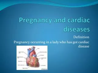
Heart –introduction • Heart, in anatomy, hollow muscular organ • it is heart shaped that pumps blood to the body. • The heart, blood, and blood vessels make up the circulatory system, which is responsible for distributing oxygen and nutrients to the body and carrying away carbon dioxide and other waste products. • The heart is the circulatory system’s power supply.
Structure of Heart • Heart valves • Heart chamber • Myocardium • Pericardium • Endocardium • Coronary arteries
Heart valves • Four valves within the heart prevent blood from flowing backward in the heart. The valves open easily in the direction of blood flow, but when blood pushes against the valves in the opposite direction, the valves close. Two valves, known as atrioventricular valves, are located between the atria and ventricles. • The other two heart valves are located between the ventricles and arteries. They are called semilunar valves
Myocardium • Muscle tissue, known as myocardium or cardiac muscle, wraps around a scaffolding of tough connective tissue to form the walls of the heart’s chambers
Pericardium • A tough, double-layered sac known as the pericardium surrounds the heart. The inner layer of the pericardium, known as the epicardium, rests directly on top of the heart muscle.
Endocardium • The inner surfaces of the heart’s chambers are lined with a thin sheet of shiny, white tissue known as the endocardium. The same type of tissue, more broadly referred to as endothelium, also lines the body’s blood vessels, forming one continuous lining throughout the circulatory system.
Coronary arteries • The heart is nourished not by the blood passing through its chambers but by a specialized network of blood vessels. Known as the coronary arteries, these blood vessels encircle the heart like a crown. • Three main coronary arteries—the right, the left circumflex, and the left anterior descending—nourish different regions of the heart muscle.
FUNCTION OF THE HEART The heart’s duties are much broader than simply pumping blood continuously throughout life. The heart must also respond to changes in the body’s demand for oxygen. • Cardiac cycle • Heartbeat • Heart sound • Cardiac output
Heart Sound • Introduction: -The mechanical activities of the heart during each cardiac cycle cause the production of the some sounds, which are called Heart Sounds. The factors involved in the production of heart sounds are as follows: -· The movements of blood through the chamber of the heart. · The movements of cardiac muscle. The movements of the valves of the heart.
ELECTROCARDIOGRAM ECG is a linear graph of the voltage fluctuation produces by the myocardium. The heart muscle posses the property of automatic rhythmic contraction, the impulse that arise in the conduction system spread through out myocardium resulting in the excitation of the muscle fibers. • This result in weak electric current which spread through the entire body. This can be recorded by placing electrode at various positions on the body and connecting them two end electro cardiac tropic apparatus.
Abnormalities • Absence of p wave increase of atrial fibrillation or nodal rhythm, sino atrial block & hypertension. • Inverted in lead I (dextocardia) • Incorrect electrode placement • Wide and notched due to left atrial enlargement • Tall and packed due to right atrial enlargement
QRS Complex • It is produced by ventricular activation on depolarization. • Q wave - -ve deflection that receives the R waves. It denotes the depolarization of ventricular septum from left to right. • R wave is the 1st +ve deflection of QRS complex it denotes the depolarization of ventricles. • S wave –v e deflection, which follows the R wave. It occurs due to depolarization of the postero basal part of the left ventricle.
Heart diseases • The arteries that nourish the heart become narrowed and unable to supply enough blood and oxygen to the heart muscle. • However, many other problems can also affect the heart, including congenital defects (physical abnormalities that are present at birth), malfunction of the heart valves, and abnormal heart rhythms. • Any type of heart disease may eventually result in heart failure, in which a weakened heart is unable to pump sufficient blood to the body.
Major causes of heart diseases Emotion Physical excitement Fatty diet Stressful life
Coronary Heart Disease • Coronary heart disease, the most common type of heart disease in most industrialized countries, • It is caused by atherosclerosis, the buildup of fatty material called plaque on the inside of the coronary arteries • Over the course of many years, this plaque narrows the arteries so that less blood can flow through them and less oxygen reaches the heart muscle.
The most common symptom of coronary heart disease is • Angina pectoris • a squeezing • chest pain that may radiate to the neck, jaw, back, and left arm. • Angina pectoris is a signal that blood flow to the heart muscle falls short when extra work is required from the heart muscle. • An attack of angina is typically triggered by exercise or other physical exertion, or by strong emotions. • Coronary heart disease can also lead to a heart attack, which usually develops when a blood clot forms at the site of a plaque and severely reduces or completely stops the flow of blood to a part of the heart.
In a heart attack, also known as myocardial infarction, part of the heart muscle dies because it is deprived of oxygen. This oxygen deprivation also causes the crushing chest pain characteristic of a heart attack. • Other symptoms of a heart attack include nausea, vomiting, and profuse sweating. About one-third of heart attacks are fatal, but patients who seek immediate medical attention when symptoms of a heart attack develop have a good chance of surviving
Arteriosclerosis • Arteriosclerosis, a group of disorders of the arteries, the tubular vessels that carry oxygen-carrying blood from the heart to the body’s organs and tissues. • In arteriosclerosis, the walls of the arteries thicken, harden, and lose their elasticity. The blood vessel channels develop twists and turns and become narrowed so that the heart must work harder than normal to pump blood through the arteries. In the disease’s advanced stage, there is a risk of a decrease in blood flow and oxygen supply to all parts of the body.
The most common form of arteriosclerosis is atherosclerosis, also known as coronary artery disease. In this condition, deposits of plaque—a material rich in greasy compounds called lipids, including cholesterol—form on the inner walls of the arteries. These deposits narrow the arterial channels and partly block the normal flow of blood through them.
The symptoms of arteriosclerosis depend upon the arteries affected. A decrease in the flow of blood through the coronary arteries, resulting in a shortage of oxygen going to the heart muscle, causes chest pains, a condition called angina pectoris. • If a blood clot forms in a coronary artery, the interruption of the blood flow can result in the death of part of the heart muscle, causing the crushing chest pains of a heart attack. • A chronic decrease in the circulation of blood to the heart may result in heart failure, which is the inability of the heart muscle to pump enough blood for the body’s requirements. Unless treated, this condition is fatal.
ISCHAEMIC HEART DISEASE Ischeimcs heart disease (IHD) is defined as acute or chronic form of cardiac disability arising from imbalance between the myocardial supply and demand for oxygenated blood. Since narrowing or obstruction of the coronary arterial system is the most common cause of myocardial anoxia, the alternate term coronary artery disease (CAD)' is used synonymously with IHD. IHD 0) CAD is the leading cause of death in most industrialised countries (about one-third of all deaths) and somewhat low incidence is observed in the developing countries. Men develop IHD earlier than women and death rates are also slightly higher for men than for women until the menopause.
ETIOPATHOGENESIS IHD is invariably cause by disease affecting the coronary arteries, the most prevalent being arteriosclerosis accounting for more than 90% cases, while other causes are responsible for less than 10% cases 01 IHD. Therefore, it is convenient to consider the etiology of IHD under three broad headings: 1. Coronary atherosclerosis 2. Superadded changes in coronary atherosclerosis 3. Non-atherosclerotic causes.
Coronary Atherosclerosis Coronary atherosclerosis resulting in 'fixed' obstruction is the major cause of IHD in more than 90% cases. Here, a brief account of the pathology of lesions in atherosclerotic coronary artery disease is presented. 1. Distribution. Atherosclerotic lesions in coronary arteries are distributed in one or more of the three major coronary arterial trunks, the highest incidence being in the anterior descending branch of the left coronary, followed in decreasing frequency, by: the right coronary artery and stil1.1ess in circumflex branch of the left coronary. About one-third of cases have single-vessel disease, most often left anterior descending arterial involvement; another one-third have two-vessel disease, and the remainder have three major vessel disease.
2. Location. Almost all adults’ show atherosclerotic plaques scattered throughout the coronary arterial system. However, significant stenotic lesions that may produce chronic myocardial ischaemia show more than 75% (three-fourth) reduction in the cross-sectional area of a coronary artery or its branch. The area of severest - involvement is about 3 to 4 cm from the coronary ostia, more often at or near the bifurcation of the arteries, suggesting the role of haemodynamic forces in atherogenesis.
3. Fixed atherosclerotic plaques. The atherosclerotic plaques in the coronaries are more often eccentrically located bulging into the lumen from one side. Occasionally, there may be concentric thickening of the wall of the artery. Atherosclerosis produces gradual luminal narrowing that may eventually lead to 'fixed' coronary obstruction. The general features of atheromas of coronary arteries are similar to those affecting elsewhere in the body and may develop similar complications like calcificatiQl1, coronary thrombosis, ulceration haemorrage, rupture and aneurysm formation.
ANGINA PECTORIS • Angina pectoris is a clinical syndrome of IHD resulting from transient myocardial ischaemia. It is characterised by paraxysmal pain in the substernal or precardial region of the chest which is aggravated by an increase in the demand of the heart and relieved by a decrease in the work of the heart. Often, the pain radiates to the left arm, neck, jaw or right arm. • There are 3 overlapping clinical patterns of angina pectoris with some differences in their pathogenesis: • Stable or typical angina • Prinzmetal's variant angina • Unstable or crescendo angina
SHOCK • Shock is complex clinical syndrome in which the circulatory system fails to maintain cellular perfusion and function CAUSATION • Hypovolaemia (decreased circulating blood volume ) • Cardiogenic shock (decreased pump function of heart) • Distributive shock(relative Hypovolaemia due to vasodialatation)
Clinical features • Fast and thready pulse • Severe fall in B.P. • Cold clammy hand and feets • Fast shallow breathing • Confusion, loss of consciousness • Renal shut down(oliguria/anuria) • Multiorgan failure
Mitral stenosis • The mitral valve is most frequently damaged by rheumatic carditis and mitral stenosis is most frequent valvular lesion of established RHD CLINICAL FEATURES • Dyspnoea • Palpitation • Pulsus parvus • Mitral facies • Taping apex beat , diastolic thrill • Loud first sound • ECG-LA enlargement
ACUTE PULMONARY OEDEMA • Transudation of fluid from pulmonary capillaries into the alveoli result in pulmonary capillaries into the perivascular space but not to the alveoli causes interstitial pulmonary oedema, a step prior to the development of pulmonary oedema
CLINICAL FEATURES The patient is • Dyspnoeic & restless • Anxious • Cyanosed & profusely sweating • Respiration is rapid • Blood pressure is raised
MYOCARDIAL DISEASE • There are two blood groups of myocardial diseases • Myocarditis Inflammation of heart muscles is called myocarditis • Cardiomyopathy Non-inflammatory myocardium involvement with unknown (primary) or known (secondary) etiology
HEART FAILURE It is the patho-physiologic state in which impure cardiac function is unable to maintain an adequate circulation for the metabolic needs of the tissue of the body. CLINICAL FEATURES • Dyspnoeic & orthoponeic • Fast & low volume pulse, pulsus alternans in LVF • Cold hands and feet with peripheral cyanosis of nails and lips • Raised JVP, positive abdomino-jugular reflux
Cardiac enlargement (apex beat shifted down and out) • RV hypertrophy seen as left parasternal and epigastric pulsation • Percussion confirms cardiac • auscultation -1st sound variable, pulmonary component of 2nd sound loud, 3rd and 4th sound may be audible.
Specialised diagnostic studies may be benefit in management of the patient such as • M.R.I. • Ultrasonography for abdominal aortic aneurysm • Cardiac catheterisation in the case of cardiogenic shock, massive pulmonary embolus • Echocardiography for VSD. Pericardial effusion
2. Rest – • reduces the demand on the heart • Adequate rest reduces venous pressure and pulmonary congestion 3. Diet – • Obese patient require a low calorie diet • Fried food are avoided • Fat is reduced • Protein content is kept normal(50-70gm) • Sodium content should not exceed 6gm of salt • Vitamin supplement may be required
ACONITUM NAPELLUS • Palpitation of the heart in young • Growing person and plethoric individuals • Congestion to both heart & lungs • Palpitation with anxiety • Cardiac oppression & even syncope • Fear of death • Hyperaemia preceding endocarditis • Confused & nervous in crowd,raises blood from least excitement
APIS MELLIFICA • Cardiac inflammation & dropsy • Sudden lancinating ,darting or stinging pain just below the heart, soon extending daigonally towards the right chest • Grat feeling of suffocation, its seems he would smother for want of air • Oedema or sudden mucus swelling • Dysnoea fidgety restlessness and anxiety, blowing sound with the diastole • Pericarditis and hydropericardium • Pulse not steady, irregular, intermitting every 3rd or 4th beat
APOCYNUM CANNABINUM • Hydropericardium, heart’s action scarcely perceptible, face bloated and anxious looking can hardly speak for want of breathe • Great dysnoea, wheezing breathing cough • pulse slow, small, irregular • General dropsy • Urine scanty
Arsenicum album • Cardiac cachexia • Endocariditis, pericaditis with restlessness • Irritable heart, trembling, irregular action of heart, intermitting • Palpitation with anguish • Valvular disease with intermittent pulse, dysnoea, anasarca • Hydrothorax and hydropericardium, with spells of suffocation
AURUM METALLICUM • Pure cardiac hypertrophy without dilatation, with increased force of heart stroke and hyperaemia of lungs • Endocarditis with loud endocardial bruits of fluttering action of heart or sudden jerks through the heart • there is violent palpitation and anxiety with congestion of blood to head and chest after exertion • Pain in heart region extends down the left arm to fingers • Pulse is small feeble but rapid and irregular
CACTUS GRANDIFLORUS • Snaguinous congestion to chest • Endocarditis and pericarditis • Sensation of constriction of heart, as if it were compressed or squeezed by a hand • Violent constriction of heart muscle, throwing the blood with great force into the aorta • Enlargement of left ventricle with grat irregularity of heart’s action • Pulse is quick, throbbing, tense and hard • Endocardial murmurs