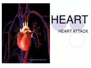
Heart
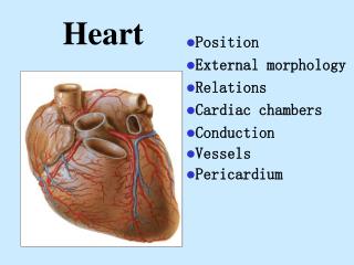
Heart. Position External morphology Relations Cardiac chambers Conduction Vessels Pericardium. Position Lies within the pericardium in middle mediastinum Behind the body of sternum and coastal cartilages 2 to 6 In front of thoracic vertebrae 5 to 8

Heart
E N D
Presentation Transcript
Heart • Position • External morphology • Relations • Cardiac chambers • Conduction • Vessels • Pericardium
Position • Lies within the pericardium in middle mediastinum • Behind the body of sternum and coastal cartilages 2 to 6 • In front of thoracic vertebrae 5 to 8 • A third of it lies to the right of median plan and 2/3 to the left
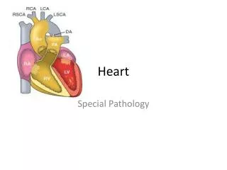
External morphology one apex, one base, two surfaces three borders three grooves
the apex formed by the left ventricle the base faces backward,upward and to the right. Formed mainly by the left atrium and partly by the right atrium. two surfaces: sternocostal and diaphragmatic surfaces three borders: right,left and inferior borders *the cardiac apecal incisure is on the inferior border and right to the apex *atrioventricular crux is a point of the coronary and posterior interventricular grooves. It is a important superficial mark three grooves: coronary groove ant. interventricular groove post. interventricular groove
Chambers • right atrium • right ventricle • left atrium • left ventricle
—two parts(divided by the terminal crest) ant.part atrium proper post.part sinus *four openings orifice of the sup.vena cava orifice of the inf.vena cava inlet orifice of the coronary sinus the right atrioventricular orifice---outlet *the fossa ovalis (oval foramen) *the triangle of Kock (tricuspid valves , orifice of the coronary sinus, tendon of todaro) Atrioventricular node is located in.
Right ventricle • inflow tracts • outflow tracts (divided by the supraventricular crest) • pulmonary valve • tricuspid valves • chordae tendineae • papillary muscles(3 groups)
Left atrium left auricle —five openings *four orifices of the pulmonary veins *the left atrioventricular orifice
Left ventricle inflow and outflow tracts (divided by the ant.cusp of the bicuspid valves) ※the papillary muscles(2 groups)
structure the walls of the heart 3 layers —endocardium *continue with the lining of the large blood vessels —myocardium *2 kinds: the ordinary cardiac muscles the specially m. — epicardium • Septum interatrial septum ----Oval fossa interventricular septum ----Membranous part
Conduction System It is consists of the special cardiac muscles 5 parts: sinoatrial node atrioventricular node, atrio-ventricular bundle Left and right branches Purkinje fibers.
—The sinoatrial node(SAN) *It is lies the junction between the right auricle and the sup.vena cava —The atrioventricular node(AVN) *It is lies in the lower portion of the interatrial septum just above the orifice of the coronary sinus —Purkinje fibers
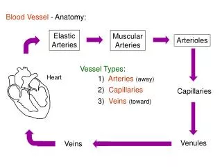
The vessels of the heart The arteries —the right coronary a. *arise from the right aortic sinus and runs along the right portion of the coronary groove *2 branches: post.interventricular branch post.branch of the left venticle *To supply right atrium,right ventricle post.1/3 of the interventricular septum,the diaphragmatic surface of left ventricle,the SAN and AVN. —The left coronary a. *arises from the left aortic sinus *2 branches: ant.interventricular br. circumflex br. *To supply the left atrium,left ventricle,the ant. surface of the right ventricle,ant.2/3 of the interventricular septum,sometimes, supply the SAN and AVN.
2. The veins of the heart—The coronary sinus *It lies in the post.portion of the coronary sulcus between the left atrium and left ventricle *It opens into the right atrium *It receives:**the great cardiac v. **the middle cardiac v. **small cardiac v.—the other veins opening into the right atrium directly *the ant. cardiac v. *the smallest v.
Pericardium 1. It comprises 2 sacs: —the fibrous pericardium —the serous pericardium: *parietal layer *visceral layer 2.The pericardial cavity: —the transverse sinus —the oblique sinus
Pericardium sinus • Transverse sinus of pericardium-posterior to ascending aorta and pulmonary trunk, anterior to superior vena cava and left atrium. • Oblique sinus of pericardium-cul-de-sac , posterior to heart, bounded by pulmonary veins on either side