Heart
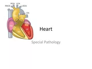
Heart. 1. general A) located within mediastinum, within the pericardial cavity B) about the size of a fist C) cone-shaped – apex facing left hip D) main function is to pump blood. Heart. 2. coverings A) surrounded by the pericardium – dual-walled structure 1) fibrous pericardium

Heart
E N D
Presentation Transcript
Heart 1. general A) located within mediastinum, within the pericardial cavity B) about the size of a fist C) cone-shaped – apex facing left hip D) main function is to pump blood
Heart 2. coverings A) surrounded by the pericardium – dual-walled structure 1) fibrous pericardium a) protects heart b) anchors it to surrounding structures c) prevents overfilling
Heart 2) serous pericardium a) 2 layers i) parietal layer (a) attached to fibrous pericardium ii) visceral layer (epicardium) (a) integral part of the heart wall
Heart b) pericardial cavity i) separates parietal and visceral layers ii) filled with pericardial fluid; creates friction-free work area 3. layers of the heart wall A) epicardium (visceral layer of the serous pericardium) 1) composed of a thin layer of CT
Heart B) myocardium 1) composed of cardiac muscle tissue C) endocardium 1) composed of simple squamous epithelium 2) is continuous with blood vessels entering & leaving heart
Heart 4. chambers A) atria 1) auricle – exterior extruding surface 2) R & L are separated by the interatrial septum a) fossa ovalis – shallow depression found in right atrium; remnant of foramen ovale
Heart 3) thin-walled – not much contracting 4) receive blood from veins a) right atrium i) superior vena cava – from structures above diaphragm ii) inferior vena cava – from structures below diaphragm iii) coronary sinus – from heart itself
Heart b) left atrium i) 4 pulmonary veins – from the lungs B) ventricles 1) separated from atria by the atrioventricular septum 2) R & L separated by interventricular septum
Heart 3) within the ventricles, 2 distinct muscle formations exist a) trabeculae carneae i) internal ridges b) papillary muscles i) finger-like projections 4) atrioventricular groove 5) interventricular sulcus
Heart C) heart valves 1) atrioventricular (AV) valves a) found between atria & ventricles b) name refers to the number of cusps (flaps) i) tricuspid valve – right side ii) bicuspid/mitral valve – left side
Heart c) attached to papillary muscles via chordae tendineae i) prevents backflow of blood into atria d) remain open when ventricles are relaxed 2) semilunar valves a) found between ventricle & its corresponding artery
Heart b) named according to the corresponding artery i) pulmonary semilunar valve – right side ii) aortic semilunar valve – left side c) remain closed when ventricles are relaxed
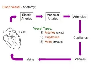
Heart 5. pulmonary circulation – by right side of heart A) de-oxygenated blood moves from the right atrium to right ventricle thru tricuspid valve B) right ventricle into pulmonary trunk thru the pulmonary semilunar valve C) to lungs for gas exchange 1) occurs in the alveoli – O2 in and CO2 out D) oxygenated blood moves back to left atrium via pulmonary veins
Heart 6. systemic circulation – by left side of heart A) oxygenated blood moves from the left atrium to left ventricle thru bicuspid/mitral valve B) left ventricle into aorta thru aortic semilunar valve C) to body for gas exchange
Heart 1) occurs in the capillaries within the tissues – O2 out and CO2 in D) de-oxygenated blood moves back to right atrium via inferior & superior vena cava 7. coronary circulation – branch of systemic loop A) aorta receives blood from L ventricle B) R & L coronary arteries receive blood from the aorta
Heart 1) right coronary artery a) supplies right atrium b) 2 main branches i) marginal artery (a) supplies anterior & lateral portions of the right ventricle ii) posterior interventricular artery (a) supplies posterior side of both ventricles
Heart 2) left coronary artery a) 2 main branches i) anterior interventricular artery (a) supplies anterior side of both ventricles ii) circumflex artery (a) supplies left atrium and portions of the left ventricle
Heart C) myocardial capillaries – site of gas exchange D) cardiac veins 1) great cardiac vein a) drains the anterior aspect of the heart 2) posterior, middle, and small cardiac veins a) drain the posterior & lateral aspects of the heart E) coronary sinus 1) empties into right atrium
Heart 8. Cardiac Muscle Contraction A) involves autorhythmic cells and cardiac muscle cells 1) autorhythmic cells a) make up the conduction system b) responsible for AP generation i) cells have an unstable resting potential
Heart ii) hyperpolarization at the end of an AP causes a closing of K+ channels and an opening of slow Na+ channels = causes movement towards threshold iii) at threshold, voltage-gated Ca++ channels open = depolarization iv) at peak, Ca++ channels close and K+ channels open = repolarization
Heart d) Conduction Pathway i) sinoatrial (SA) node (a) considered normal pacemaker (i) 90-100 AP’s/min = 90-100 b.p.m.
Heart (b) under control of nervous and endocrine systems (i) at rest produces about 75 AP’s/min = 75 b.p.m. (c) impulse travels to AV node via internodal pathway (d) impulse also travels to atrial myocardium via gap junctions (intercalated discs) (i) causes atrial contraction
Heart ii) atrioventricular (AV) node (a) AV nodal delay (i) 0.1 sec (ii) allows for complete atrial contraction (ventricular filling) (b) under nervous & endocrine control
Heart iii) bundle of His (AV bundle) (a) electrically connects atria & ventricles iv) left & right bundle branches (a) left and right sides of heart
Heart v) Purkinje fibers (a) start near the apex & moves up thru ventricles (b) site of synapse between conduction system & ventricular myocardium
Heart 2) cardiac muscle cells a) striated, branching & mononucleated b) intercalated discs – cellular junctions that allow ion transport between cells i) allows heart to act as a single, coordinated, functional unit
Heart ii) longer refractory period than skeletal muscle tissue; cannot undergo tetanus c) AP generation i) depolarization – opening of voltage-gated Na+ channels ii) repolarization – opening of voltage-gated K+ channels
Heart iii) plateau – opening of voltage-gated Ca++ channels, leakage of K+
Heart 3) Process of Contraction a) AP generated in SA node travels to atrial myocardium and AV node i) causes atrial contraction b) AP travels from AV node to bundle of His then along bundle branches to the Purkinje fibers i) Purkinje fibers synapse with the ventricular myocardium
Heart c) AP travels down the sarcolemma and causes voltage-gated Ca++ channels in sarcolemma to open d) Ca++ moves into the cell from the ECF e) This causes an opening of Ca++ channels in the SR, causing larger amounts of Ca++ to be released = calcium-induced calcium release (20% from ECF & 80% from SR) f) Ca++ binds to troponin...sliding of filaments
Heart 9. Cardiac Cycle A) Series of events occurring during one heartbeat; 4 events occur 1) atrial & ventricular systole 2) atrial & ventricular diastole B) 3 phases 1) relaxation period a) occurs just after blood is ejected from the ventricles
Heart b) semilunar valves are open & AV valves are closed c) characterized by: i) ventricular diastole (a) causes decreased ventricular P ii) closing of semilunar valves (a) causes “dup” sound iii) opening of AV valves
Heart 2) ventricular filling a) begins when AV valves open b) characterized by: i) rapid ventricular filling (80%) ii) atrial contraction (20%) c) end diastolic volume (EDV) i) volume of blood in the ventricle just prior to contraction
Heart 3) ventricular ejection a) characterized by: i) ventricular systole (a) causes increased ventricular P ii) closing of AV valves (a) causes “lub” sound
Heart iii) opening of semilunar valves iv) ventricular ejection (a) stroke volume (~70ml) v) atrial filling also occurs during this phase
Heart 10. Cardiac Output – total amount of blood pumped by each ventricle per minute A) CO = SV x HR (5.25L/min) B) Regulation of Cardiac Output – 2 mechanisms 1) Regulation of Stroke Volume – 3 factors a) preload – stretch on the cardiac muscle just before contraction i) associated with EDV – end diastolic volume ii) Frank-Starling Law of the Heart
Heart b) contractility – strength of contraction i) positive inotropic agent (a) promote Ca++ movement into cells ii) negative inotropic agent (a) inhibit Ca++ movement into cells c) afterload – pressure the ventricles must overcome to eject blood
Heart 2) Regulation of HR a) ANS Control i) Cardiovascular center (a) Composed of 3 centers (i) Cardioacceleratory center (ii) Cardioinhibitory center (iii) Vasomotor center
Heart (b) receives input from: (i) chemoreceptors in aortic arch & bifurcation of common carotid artery (ii) baroreceptors in aortic arch and carotid sinus (iii) proprioceptors in skeletal muscles & joints (c) send output signals via:
Heart (i) sympathetic NS (responds to hypoxia, hypercapnia, acidosis or low BP) (a) stimulates cardiac accelerator nerves (NE) (i) innervate the SA & AV nodes (ii) also innervate the ventricular myocardium (ii) parasympathetic NS (responds to high BP) (a) stimulates the Vagus nerve (ACh) (i) innervates the SA & AV nodes
Heart b) Hormonal Control (low BP) i) epinephrine & norepinephrine c) Other Factors i) hypernatremia – blocks Ca++ movement into SA node ii) hyperkalemia – inhibits AP generation iii) hypercalcemia – increases conc. gradient iv) hypocalcemia – decreases conc. gradient
Heart 11. Electrocardiogram (ECG or EKG)
Heart A) P-wave 1) atrial depolarization B) QRS-complex 1) ventricular depolarization 2) atrial repolarization is occurring but is masked C) T-wave 1) ventricular repolarization
Heart 12. Heart Disorders A) Valve disorders 1) Heart murmur – abnormal heart sounds a) Stenosis – valve flaps become stiff and narrowed thereby restricting normal blood flow b) Incompetent valve – valves fail to close properly resulting in a backflow of blood
Heart c) Mitral valve prolapse (MVP) – chordae tendineae are abnormal and/or the papillary muscle malfunction resulting in the flaps becoming inverted
Heart B) Arrhythmias – abnormal heart rate 1) Tachycardia – more than 100 beats per minute a) may be caused by elevated temp, certain drugs, stress, or heart disease
Heart 2) Bradycardia – less than 60 beats per minute a) may be caused by low temp, certain drugs, or parasympathetic activation 3) Fibrillation – uncoordinated or quivering heartbeat a) caused by damage/defect of conduction system
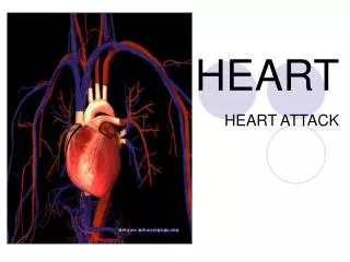
Heart 4) Heart block – inability of impulse to reach ventricles a) blockage in the AV node, bundle of His or one of the bundle branches C) Others 1) Myocardial Infarction (MI) – “heart attack” a) Infarction – tissue death due to loss of blood supply