Download

1 / 35
350 likes | 444 Views
Explore the intricate pathways of blood through the heart and lungs, major vessels involved, functions of ventricles, atrioventricular and semilunar heart valves, coronary arteries, cardiac muscle structure, and the role of blood pressure and resistance in circulation. Learn about EKG importance and monitoring heart health.

E N D
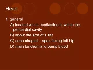
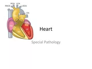
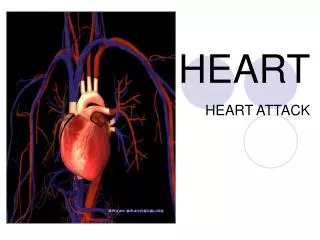
Heart • Considered to be a duel pump because the both sides work independently of one another. • The right side pumps blood to the lungs. • (pulmonary circulation) • The left side pumps blood to all of the other organs of the body • (systemic circulation)
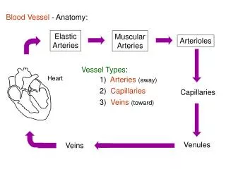
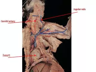
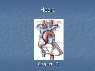
Major Vessels of the Heart • Vessels returning blood to the heart include: • Superior and inferior venae cavae • Right and left pulmonary veins • Vessels carrying blood away from the heart include: • Pulmonary trunk, which splits into right and left pulmonary arteries • Ascending aorta (three branches) –, left common carotid, and subclavian arteries
Ventricles of the Heart • Ventricles eject blood from the heart • Right ventricle pumps blood into the pulmonary trunk which will go to the lungs. • Left ventricle pumps blood into the aorta which will go to the rest of the body
Atrioventricular Heart Valves • Heart valves ensure unidirectional blood flow through the heart. • Atrioventricular (AV) valves lie between the atria and the ventricles. • Tricuspid Right • Bicuspid (Mitral) Left • AV valves prevent backflow of blood into the atria when ventricles contract. • Chordae tendineae anchor AV valves to papillary muscles • First heart sound (LUB) when valves close
During diastole there is less pressure in the in ventricle. AV Valves open and filling the ventricle. • During systole the AV valves prevent the back flow of blood into the atrium. • Failure to prevent the blood from going back into the atria (systolic heart murmur)
Semilunar Heart Valves • Aortic semilunar valve lies between the left ventricle and the aorta. • Pulmonary semilunar valve lies between the right ventricle and pulmonary trunk • Semilunar valves prevent backflow of blood into the ventricles. • Second heart sound (DUB) when valves close.
Figure 19.9b • During ventricular systole the semilunar valves allow blood to be ejected from the ventricles into the aorta and pulmonary trunk. • During diastole they prevent the back flow of blood back into the ventricles. • Failure to prevent the backflow of blood into the ventricles. (diastolic murmur)
Coronary Arteries • Left coronary artery (LCA) • anterior interventricular branch • supplies blood to interventricular septum and anterior walls of ventricles • circumflex branch • passes around left side of heart in coronary sulcus, supplies left atrium and posterior wall of left ventricle • Right coronary artery (RCA) • right marginal branch • supplies lateral R atrium and ventricle • posterior interventricular branch • supplies posterior walls of ventricles
Cardiac muscle is short and striated. • Ability to beat independent of stimulation from the nervous system. • ANS • Functional syncytium: • intercalated discs connect cardiac cells which allow free passage of ions. • This allows the spread of action potentials from one myocyte to another. • The result is a coordinated contraction that moves the blood out of the heart.
Blood Pressure (BP) • Force per unit area exerted on the wall of a blood vessel by its contained blood. • Expressed in millimeters of mercury (mm Hg) • Measured in reference to systemic arterial BP in large arteries near the heart. • The differences in BP within the vascular system provide the driving force that keeps blood moving from higher to lower pressure areas.
Resistance • Resistance – opposition to flow • Measure of the amount of friction blood encounters as it passes through vessels. • Generally encountered in the systemic circulation. • Referred to as peripheral resistance (PR) • The three important sources of resistance are blood viscosity, total blood vessel length, and blood vessel diameter.
Resistance Factors: Viscosity and Vessel Length • Resistance factors that remain relatively constant are: • Blood viscosity – thickness or “stickiness” of the blood the greater the resistance. • Blood vessel length – the longer the vessel, the greater the resistance encountered. • Diameter- Smaller diameter arterioles are the major determinants of peripheral resistance • Fatty plaques from atherosclerosis: decreases both diameter and elasticity of vessel which results in: • Cause turbulent blood flow • Dramatically increase resistance due to turbulence
Blood Flow, Blood Pressure, and Resistance • Blood flow (F) is directly proportional to the difference in blood pressure • If P increases, blood flow speeds up; if P decreases, blood flow declines • Blood flow is inversely proportional to resistance (R) • If R increases, blood flow decreases • R is more important than P in influencing local blood pressure.
Why Get an EKG • Unexplained chest pain, or reduced blood flow to the heart (ischemia), shortness of breath, dizziness, fainting, or rapid and irregular heartbeats (palpitations). • Identify ventricle hypertrophy and other changes of the myocardium. • Check how well mechanical devices, such as pacemakers or defibrillators implanted in the heart, are working to control a normal heartbeat.
Electrocardiography • Electrical activity is recorded by electrocardiogram (ECG) • P wave corresponds to depolarization of SA node resulting in atrial systole • QRS complex corresponds to ventricular depolarization resulting in Ventricular contraction and blood being ejected from the heart. • Atrial repolarization is hidden with in QRS complex • ST segment - ventricular systole • T wave corresponds to ventricular repolarization
Normal Sinus Rhythm • The heart is being paced by the SA node. There is a degree of regularity between all components of EKG • Normal heart rate ranges between 60-100bpm. • 75bpm( average) dependent on activity. • Find a QRS that falls on a solid black line. • Then count 300-150-100-75-60-50-43 for each successive black line. Distance between QRS complexes are 5 black lines =60 Beats per minute
Sinus Bradycardia (SB) is defined as a sinus rhythm with a rate below 60 bpm. • Normally found in well trained persons and during sleep. • May also be found in patients post myocardial infarction • Beta Blockers reduce sympathetic input to the heart reducing both cardiac workload and blood pressure. • If on this medication heart rate is not a valid measure of exertion.
Tachycardia • Defined as a heart rate greater than 100bpm • This is a perfectly normal rhythm if you are exercising. • Heart beats faster under the influence of the SNS in order to meet the bodies increased demand of oxygen. • Stress, anxiety, or underline pathology may result in an elevated HR. • Context is important.
Atrial Fibrillation • Multiple ectopic foci fire chaotically in the atrium • This diminished the atriums ability to contract and results in pooling in the atrium. • (High risk of blood clot formation =need blood thinners) • Loss of atrial kick = reduced SV • May lead to a more severe arrhythmia.
Premature Ventricular Contraction (PVC) Ventricles contract before the atria in a cardiac cycle The SA node is not pacing the heart here. • typically caused by emotional stress, lack of sleep, smoking or stimulants (caffeine) which initiate an AP in the ventricles
Ventricular Tachycardia( V-Tach) • Heart rate greater then 150 that originates from an irritable foci in the ventricle. • It is a regular rhythm but is hemo-dynamically inefficient. The heart CO will be very poor due to reduced ventricular filling. • Reduced CO leads to poor coronary perfusion. • Will lead to Ventricular Fibrillation
Ventricular Fibrillation( V-FIB) • This is the most dangerous rhythm. There is no discernable pattern. • Multiple areas of the ventricles are initiating impulses at the same time resulting in a quivering heart instead of a contracting heart. • The heart will not efficiently fill up with blood. • Oxygen will not be delivered to the tissues. • Toe tag is eminent unless a defibrillator ( AED) is near.