Download

1 / 47
480 likes | 776 Views
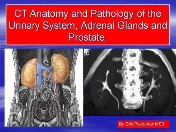
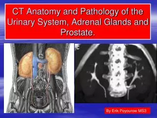
Pathology of the prostate. KVB. Anatomy of the prostate. Retroperitoneal organ encircling the neck of the urinary bladder & urethra Devoid of a distinct capsule In normal adult, weighs approximately 20g Can be divided into 4 biologically & anatomically distinct zones or regions.

E N D
Anatomy of the prostate • Retroperitoneal organ encircling the neck of the urinary bladder & urethra • Devoid of a distinct capsule • In normal adult, weighs approximately 20g • Can be divided into 4 biologically & anatomically distinct zones or regions
Zones of the prostate • Peripheral • Central • Transitional • Region of the anterior fibromuscular stroma Most hyperplasias arise in the transitional zones, whereas most carcinomas arise in the peripheral zone.
Histology of the prostate • Tubuloalveolar organ composed of glands which are lined by epithelium • Lining is composed of 2 layers of cells: a basal layer of low cuboidal epithelium covered by a layer of columnar secretory cells • The epithelium shows papillary infolding & rests on a basement membrane • Glands are separated by abundant fibromuscular stroma
Pathologic processes affecting the prostate: • Inflammation (prostatitis) • Benign nodular hyperplasia • Tumours
Inflammations • Acute & chronic bacterial prostatitis • Chronic abacterial prostatitis • Granulomatous prostatitis
Acute bacterial prostatitis • Caused by organisms that cause UTI: E.coli, other gram-negative rods, enterococci & staphylococci • Presents with fever, chills & dysuria • Diagnosis is by urine culture & clinical features
Morphology of acute prostatitis • Minute disseminated & coalesced abscesses , may cause enlargement of the prostate • Histologically : areas of necrosis with infiltration of neutrophils
Chronic bacterial prostatitis • Difficult to diagnose & treat • Presents with low-backache, dysuria & suprapubic discomfort • Usually due to recurrent UTI caused by same organism
This is chronic prostatitis. Numerous lymphocytes are seen in the stroma between the glands.
Granulomatous prostatitis Common causes: • Intracystic instillation of BCG for the treatment of superficial bladder cancer • Fungal infections in immunocompromised host • Reaction to the secretions released from ruptured ducts & acini Importance: • Has to be differentiated from TB cystitis • Since the prostate fells hard on rectal examination it could be mistaken for carcinoma
Nodular hyperplasia/Benign prostatic hyperplasia (BPH) • Common in men over 50 years • Pathogenesis: • Related to action of estrogens & androgens • Occurs due to altered normal ratio of estrogen: testosterone that occurs in the elderly • Estrogen sensitizes prostatic tissue to subsequent action of DHT which is derived from plasma testosterone • Mediated by Dihydrotestosterone (DHT), which is secreted by stromal cells of prostate • Once synthesized, acts in an autocrine fashion on stromal cells & in a paracrine fashion by diffusing into neighbouring epithelial cells
Pathologic features of BPH: Gross: • Prostate enlarged, nodular, firm & rubbery • Weight: 2x or more than normal • Central portion expanded by nodules that compress the peripheral part of the prostate ( forming a “surgical” or pseudocapsule) • Urethra is compressed, deformed & tortuous
Microscopy of BPH • Hyperplastic glands lined by columnar & cuboidal cells, often projecting into the lumen • 2 layers of epithelium are present • Stroma is composed of hyperplastic smooth muscle cells & fibroblasts
Benign prostatic hyperplasia involving both glands and stroma High power view of glandular hyperplasia
Clinical features of BPH • Dysuria (urge to urinate frequently,weak stream, halting, incomplete emptying of the urinary bladder) • Urinary bladder infections • Acute urinary retention which requires immediate catheterization
Complications of BPH • Cystitis • Calculi formation due to stagnation of urine & infection • Prostatitis, epididymoorchitis • Hydroureter, hydronephrosis • Acute retention of urine • Obstructive uropathy with renal failure
A frequently performed operation for symptomatic nodular prostatic hyperplasia is a transurethral resection (TURP), which yields the small "chips" of rubbery prostatic tissue seen here.
Prostatic carcinoma Key facts: • Disease of old age (over 50 years) • Uncommon among Asians, greater incidence with blacks, whites • Role of hormones: Ca prostate never develops in men castrated before puberty • Prostate specific antigen (PSA) is used for screening & monitoring of tumours • PSA is sensitive but has low specificity
Etiology • Endocrinologic factors- Androgens • Racial factors- More common in African Americans than in whites, Japanese or Chinese • Environmental factors- high fat diet, exposure to polycyclic aromatic hydrocarbons • Genetic basis- familial cases • BPH- not proved
Prostatic intraepithelial neoplasia (PIN) • Presumptive precursor lesion • Consists of benign glands with intra-acinar proliferation of cells that demonstrate nuclear anaplasia • PIN glands are surrounded by a patchy layer of basal cells & a basement membrane
This is prostatic intraepithelial neoplasia (PIN), a precancerous cellular proliferation found in a single acinus or small group of acini.
The finding of PIN suggests that prostatic adenocarcinoma may also be present
Types of carcinoma prostate • Latent carcinoma: unexpected finding in autopsy • Incidental carcinoma: in 15-20% of TURP done for BPH • Occult carcinoma: presents with features of metastases but primary is not evident • Clinical c arcinoma: detected by PR, other investigations & is symptomatic
Symptoms • By the time symptoms appear, the carcinoma, is palpable on PR as a hard mass which is fixed to surrounding tissue. • Symptoms are: • Urinary obstruction with dysuria • Retention of urine • Hematuria • Bony pain due to metastases
Pathologic features of prostatic carcinoma • Multifocal induration, most often in the outer zones & posterior lobe (can be palpated on rectal examination)
On histology, carcinoma prostate can be: • Adenocarcinoma (96% of cases) • Transitional cell carcinoma • Squamous cell carcinoma • Undifferentiated carcinoma
At the right are normal prostatic glands containing scattered corpora amylacea. At the left is prostatic adenocarcinoma. Note how the glands of the carcinoma are small and crowded.
At high magnification, the neoplastic glands of prostatic adenocarcinoma are still recognizable as glands, but there is no intervening stroma
Prominent nucleoli are seen in the nuclei of this prostatic adenocarcinoma, which is a characteristic feature.
Immunoperoxidase staining with antibody to prostate specific antigen (PSA). PSA is also used as a serum test to monitor or detect prostate cancer.
Spread of Ca prostate • Local spread: Tends to invade nerves, seminal vesicles & adjacent pelvic organs (local extension) • Lymphatic spread: To para-aortic, iliac lymph nodes • Hematogenous metastases : most often found in the vertebrae & sacrum; can also occur in kidneys,lungs & brain • Bony metastases are often osteoblastic & are associated with elevated serum alkaline phosphatase • Visceral involvement occurs rarely
Grading of prostatic carcinoma • The Gleason system is employed which stratifies prostate cancers into 5 grades based on the glandular patterns & degree of differentiation seen under low power magnification. • Grade 5 tumors show least differentiation • Gleason score is based on Gleason grading : low- grade tumours have a relatively good prognosis & high-grade tumours are invariably lethal
Staging of carcinoma prostate: TNM T1: Cancer found incidentally in TURP done for BPH T2: Organ-confined cancer T3a: Extraprostatic extension without seminal vesicle involvement T3b: Extraprostatic extension with seminal vesicle involvement T4: Direct invasion of contiguous organs
Prognosis of prostate carcinoma • Localized low-grade tumors: 95% 10-year survival rate • High-grade tumors extending to local tissues: 50% 5-year survival rate • High-grade tumors with bone metastases or spread to distant sites: poor (2-3 year survival)
Most useful techniques for diagnosis of prostate carcinoma • PSA: useful for screening & monitoring of tumours • Biopsy: Needle Bx & TURP • Ultrasound • CT scanning: can assess extent of spread
Serum markers in diagnosis & monitoring of carcinoma prostate • PSA: Normal 0-4ng/ml; >10ng/ml is diagnostic of carcinoma prostate • Prostatic acid phosphatase (PAP): elevated levels found in prostatic cancer that has extended beyond the capsule or which has metastasized.