Download

1 / 23
270 likes | 414 Views
Dive deep into the anatomy, epidemiology, and histopathologic features of prostatic hyperplasia and cancer. Learn about BPH pathogenesis and clinical features, and explore the gross morphology and microscopy of this condition.

E N D
Prostate Pathology Sufia Husain. Pathology Department KSU, Riyadh April 2017 Reference: Robbins & Cotran Pathology and Rubin’s Pathology
Lecture Outline: Prostatic hyperplasia and cancer of the prostate. At the end of this lecture, the student should be able to: • Understand the basic anatomical relations and zones of the prostatic gland. • Know the epidemiology, pathogenesis and histopathologic features of benign prostatic hyperplasia and carcinoma of the prostate.
http://napaurology.com http://www.yoursurgery.com
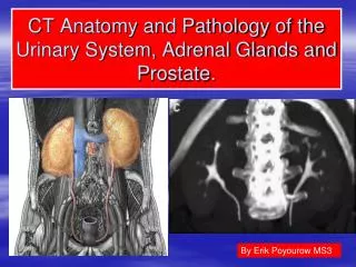
Prostate anatomy • Prostate weighs 20 grams in normal adult • Retroperitoneal organ, encircling the neck of bladder and urethra • Devoid of a distinct capsule • The prostate is divided into different zones. They are Central, Peripheral and Transitional zones. The transition zone is the middle area of the prostate, between the peripheral and central zones. It surrounds the urethra as it passes through the prostate • The majority of prostate cancers are found in the peripheral zone and benign nodular hyperplasia in the transitional zone. • Microscopically the prostate is a tubulo-alveolar organ. The prostate glands are lined by two layers of cells, basal cells and columnar secretory cells http://www.theprostatecentre.com
Normal prostate Picture Taken from Robbins and Cotran Pathologic basis of disease. 8th edition, Chapter 21, 2010 Sanders
Benign Prostatic Hyperplasia (BPH) • Also known as benign nodular hyperplasia • Extremely common lesion in men over age 50. • 20% in men over age 40, up to 70% by age 60, and 90% by age 80. • Hyperplasia of glands and stroma results in large nodular enlargement in the periurethral region of the prostate. • Once the nodules become large they compress the prostatic urethra causing either partial, or complete obstruction of the urethra. • Nodular hyperplasia is not a premalignant lesion
BPH: pathogenesis • The essential cause of BPH is unknown • The pathogenesis is related to the action of androgen. • Dihydrotestesterone (DHT) is the ultimate mediator for prostatic growth. It increases the proliferation of stromal cells and inhibits epithelial cell death.Therefore DHT is implicated in the pathogenesis of both benign prostatic hyperplasia (BPH) and prostate cancer. • Testosterone is converted to dihydrotestosterone (DHT) by 5-alpha reductaseenzymes. • Drugs that act as inhibitors of 5-alpha reductase, therefore have an important role in the prevention and treatment of BPH and prostate cancer. • Prepubertal castration prevents BPH
BPH: gross morphology • The prostate weighs between 60 and 100 grams • The hallmark of BPH is nodularity due to glandular and fibro-muscular proliferation. • Nodular hyperplasia begins in the inner aspect of the prostate gland, the transition zone • Cut-section shows nodules which vary in size, color and consistency depending on which element is proliferating more (glandular or fibro-muscular). • It compress the wall of the urethra resulting in a slit-like orifice. http://www.drbriffa.com
BPH: microscopy • Microscopically, the main feature of BPH is nodularity • The nodules can be: • purely stromal nodules composed mainly of fibromuscularelement OR • fibroepithelial with both glandular and fibromuscular component. There is aggregation of small to large to cystically dilated glands, lined by two layers of epithelium surrounded by fibromuscularstroma. • Needle biopsy doesn’t sample the transitional zone where BPH begins and occurs, therefore the diagnosis of BPH cannot be made on needle biopsy
BPH: clinical feature & treatment C/F: • The nodule compress the prostatic urethra and cause uretheralobstruction. This leads to retention of urine in the bladder with associated bladder hypertrophy. • The inability to empty the bladder completely leads to increase volume of residual urine and therefore infection • Increased urinary frequency • Nocturia • Difficulty in starting and stopping the stream of urine • Dysuria • Some patients present with acute urinary retention. Treatment: • Mild cases of BPH may be treated with α-blockers and 5-α-reductase inhibitors • Moderate to severe require transurethral resection of the prostate (TURP)
Nodular hyperplasia http://pixgood.com/benign-prostatic-hyperplasia-histology.html
Figure A & B: BPH Rubin's Pathology : Clinicopathologic Foundations of Medicine, 5th Edition, 2008 Lippincott Williams & Wilkins
Prostatic Adenocarcinoma • The a common form of cancer in men • Disease of men over age 50 • More prevalent among African Americans • These tumors show a wide range of clinical behaviors • Etiology: Several risk factors : Age, race, family history, hormone level (androgens) and environmental influences. • Androgen are believed to play a major role in the pathogenesis
Prostatic Adenocarcinoma: morphology • 70% arises in the peripheral zone of the posterior part of the gland • Tumor is firm and gritty and is palpable on rectal exam • Spread by direct local invasion and through blood stream and lymph • Local extension most commonly involves the periprostatic tissue, seminal vesicles and the base of the urinary bladder (leading to ureteral obstruction)
Prostatic Adenocarcinoma: microscopy • Histologically, most lesions are adenocarcinomas that produce well-defined gland patterns. • The malignant glands are lined by a single layer of cuboidal or low columnar epithelium with large nuclei and one or more large nucleoli. Nuclear pleomorphism is not marked. The outer basal cell layer typical of benign glands is absent • Commonly there is perineuralinvasion.
ProstaticAdenocarcinoma • Metastases first spread via lymphatics: initially to the obturator nodes and eventually to the para-aortic nodes • Hematogenous extension occurs chiefly to the bones. The bony metastasis are typically osteoblastic .
Prostatic Adenocarcinoma: Gleason Grading & Scoring • Gleason system is a histological grading and scoring system for prostatic adenocarcinoma done on the microscopic level. • There are five grades (1 to 5) depending on the degree and pattern of differentiation as seen microscopically (in which they range from, grade 1= well-differentiated to grade 5= very poorly differentiated). • The two most common types of grades seen in the biopsy for each cancer case are added to produce a combined Gleason score. • Gleason Grading and Scoring in prostate cancer is very useful in predicting prognosis of a patient.
ProstaticAdenocarcinoma: Staging • Staging in prostate cancer depends on the TNM system. It is the most important indicator of prognosis.
Prostatic Adenocarcinoma: clinical features • Microscopic (very small size) cancers are asymptomaticand are discovered incidentally. • Most arise in the peripheral zone, away from urethra and therefore the urinary symptoms occur late. • Occasionally patients present with back pain caused by vertebral metastases • Careful digital rectal examination may detect some early cancers. • PSA (Prostate Specific Antigen) levels are important in the diagnosis and management of prostate cancer. However, 20% - 40% of prostate confined cancers have low PSA • PSA is organ specific but not cancer specific because it could be increased in BPH and prostatitis. • A transrectal needle biopsy is required to confirm the diagnosis http://www.medicalhealthguide.com
Prostatic Adenocarcinoma: treatment • Surgery, radiotherapy and hormonal therapy • 90% of treated patients are expected to live for 15 years • Currently the most acceptable treatment for clinically localized cancer is radical surgery • Locally advanced cancers can be treated by radiotherapy and hormonal therapy. Hormonal therapy (Anti-androgen therapy) can induce remission. • Advanced, metastatic carcinoma is treated by androgen removal treatment, either by orchiectomy or by hormonal anti-androgen therapy. • The prognosis depends on the Gleason score and stage of tumor.
Prostatic intraepithelial neoplasia (PIN) • PIN is the precursor lesion for invasive carcinoma. It can be low grade PIN or high grade PIN. (high grade PIN is like carcinoma in situ) • PIN like carcinoma occurs in the peripheral zone.