Download
1 / 37
380 likes | 814 Views
Gallbladder and Biliary Tract Disease. Cholelithiasis. Cholelithiasis. Cholelithiasis is the pathologic state of stones or calculi within the gallbladder lumen . A common digestive disorder worldwide: 20 million Americans have gallstones

E N D
Cholelithiasis • Cholelithiasis is the pathologic state of stones or calculi within the gallbladder lumen. • A common digestive disorder worldwide: • 20 million Americans have gallstones • 700,000 cholecystectomys performed annually in the U.S. • Most common gastrointestinal disorder requiring hospitalization
Cholelithiasis Most gallstones are composed primarily of bile (80%); remainder are composed of a mixture of bile components Each type of stone has a particular pathophysiology and specific set of risk factors that alter the equilibrium and solubility of the components of bile. Pigment stones Cholesterol stones
Cholelithiasis Asymptomatic gallstone patients develop complications at an annual rate of 1-2%. In symptomatic patients, the complication rate increases to 3%. Manifestations of cholelithiasis: • Many persons are asymptomatic • Early symptoms are epigastic fullness after meals or mild distress • Biliary colic (if stone is blocking cystic or common bile duct): steady pain in epigastric or RUQ of abdomen lasting up to 5 hours with nausea and vomiting • Jaundice may occur if there is obstruction of common bile duct
Cholelithiasis • Sonography is the procedure of choice for identifying gallstones. • Current high-resolution, real-time ultrasound (US) can identify gallstones as small as 2 mm, with a sensitivity greater than 95%. • The technique is rapid, noninvasive, can be performed at the bedside, and does not involve ionizing radiation. • Ultrasonic Criteria for Cholelithiasis • Intraluminal brightly echogenic structure • Stones > 3mm will produce an acoustic shadow • Stones will usually seek gravitational dependency
Cholelithiasis Ultrasound Ultrasound image obtained with a 3,5-MHz transducer demonstrates the multiple stones in the gallbladder with typical acoustic shadows.
Cholelithiasis Ultrasound Ultrasound image obtained with a 3,5-MHz transducer demonstrates a stone in the gallbladder with typical acoustic shadow. NORMAL GALLBLADER
Cholelithiasis Ultrasound Ultrasound image demonstratesmultiple shadowing stones.
Cholelithiasis Ultrasound • Image Patterns: • Stones with shadowing • Stones without shadowing • Gravel • GB filled with stones • Floating stones as fluid level in bile • Adherent Gallstones • Dilation of common bile duct
Cholelithiasis Ultrasound Ultrasound image obtained with a 3-MHz transducer demonstrates pyramidal nonshadowing stones.
Cholelithiasis Ultrasound Wall-echo shadow sign indicates a stone-filled gallbladder.
Cholelithiasis Ultrasound Layer of gravel with shadowing
Cholelithiasis • Limitations of Techniques: • US: False negatives may occur with small stones in the presence of biliary sludge. The technique is operator-dependent. Inadequate visualization of the gallbladder may occur in obese or contracted patients, or in patients with abdominal wounds. • Radiographs: Only 15-20% of stones are visible on plain radiographs. • CT: Only 74-79% of gallstones are identified in patients with CT. CT is not a screening tool for uncomplicated cholelithiasis. • MRI: MRI is not a screening tool. Stones may be incidental findings on abdominal MRI.
Cholelithiasis • Only 15-20% of stones are visible on plain abdominal film.
Cholelithiasis CT Findings CT demonstrates a layer of calcific-dense material in the gallbladder that may be gravel or milk of calcium bile Noncontrast CT demonstrates a typical, laminated, calcified gallstone.
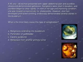
Cholelithiasis ERCP findings in cholelithiasis Gallbladder and CBD stones
Acute cholecystitis Manifestations of acute cholecystitis • Episode of biliary colic involving RUQ pain radiating to back, right scapula, or shoulder; the pain may be aggravated by movement, or deep breathing and may last 12 – 18 hours • Anorexia, nausea, and vomiting • Fever with chills
Acute cholecystitis • Sonographic features of AC include the following: • Calculi in the GB in more than 90% of patients. • Anterior GB-wall thickness of more than 3 mm • Positive Murphy sign (pain on compression of the GB with the ultrasound probe) • Pericholecystic fluid in severe cases (sign of actual or impending perforation) • Acalculous cholecystitis: Five percent of cases are not associated with gallstones. • Increased transverse GB diameter >4-5 cm • GB distension: About 93% of patients with a GB volume of greater than 70 mL have AC. • Loss of definition of GB margins
Acute cholecystitis Ultrasound Ultrasound image demonstrates: thickening of the gallbladder wall.
Acute cholecystitis Ultrasound Ultrasound image demonstrates: a calculus at the neck of the gallbladderwith acoustic shadowing and thickening of the gallbladder wall. Gallbladder is slightly enlarged
Acute cholecystitis Ultrasound Ultrasound image demonstrates: thickening of the gallbladder wall and loss of definition of GB margins.
Acute cholecystitis CT Findings Acute nongangrenous cholecystitis. CT scan shows pericholecystic fluid From: CT Findings in Acute Gangrenous Cholecystitis G. L. Bennett et al.AJR 2002; 178:275-281
Acute cholecystitis Complications of cholecystitis • Chronic cholecystitis occurs after repeated attacks of acute cholecystitis; often asymptomatic • Empyema: collection of infected fluid within gallbladder • Gangrenous cholecystitis with perforation leading to peritonitis or abscess formation • Pancreatitis, liver damage, intestinal obstruction Complications are more common in patients with small, multiple stones.
Acute cholecystitis Ultrasound Transverse and longitudinal scans demonstrate a complex echo pattern in the area of the gallbladder and pericholecystic fluid - acute gangrenous cholecystitis.
Acute cholecystitis CT Findings Acute gangrenous cholecystitis. CT scan with IV contrast material shows intraluminal linear densities corresponding to intraluminal membranes. Note lack of contrast enhancement of gallbladder wall and pericholecystic inflammation. renal cyst From: CT Findings in Acute Gangrenous Cholecystitis G. L. Bennett et al.AJR 2002; 178:275-281
Acute cholecystitis Ultrasound Acute gangrenous cholecystitis. Sonography demonstrates an anechoic fluid mass situated in the wall of the gallbladder. Sonograms shows marked laminated sonolucent thickening of the gallbladder wall, with the lumen of the gallbladder full of sludge. Gallbladder is enlarged
Acute cholecystitis CT Findings Acute gangrenous cholecystitis. CT scan with IV contrast material shows air in gallbladder lumen. From: CT Findings in Acute Gangrenous Cholecystitis G. L. Bennett et al.AJR 2002; 178:275-281
Acute cholecystitis CT Findings Abscess Acute gangrenous cholecystitis. CT scan shows loculated fluid attenuation abnormality adjacent to gallbladder, consistent with abscess (a). Defect in gallbladder wall is shown (perforation). White arrow shows pericholecystic inflammation (leading to peritonitis). From: CT Findings in Acute Gangrenous Cholecystitis G. L. Bennett et al.AJR 2002; 178:275-281
Gallbladder Sludge The term biliary sludge refers to a characteristic ultrasound picture of movable, low-amplitude echoes that layer in the most dependent part of the gallbladder and are not associated with acoustic shadowing. Sludge is composed of cholesterol crystals, calcium bilirubinate granules, and mucin glycoprotein suspended in bile and forms in an environment that combines a high mucus concentration, dysmotility, and stasis. The cholesterol and calcium bilirubinate crystals in biliary sludge can lead to gallstone formation.
Gallbladder Sludge Ultrasound Longitudinal scan through the gallbladder shows layering of sludge in the gallbladder lumen. Sludge can appear, disappear, and reappear, its formation is a dynamic, reversible process
Gallbladder Sludge Ultrasound • Clinical association with: • hyperalimentation, • hemolysis, • fasting, • pregnancy, • post-op state, • cirrhosis • Differentiate from: • hematobilia, • biliary tract tumors, • purulent bile This longitudinal view of the gallbladder, as imaged from the gastric antrum, reveals dependent echogenic sludge.
Gallbladder polyps • It is estimated that as many as 4% of gallbladders examined by ultrasound will have evidence of polyp formation. • The 95% of all gallbladder polyps do not give rise to cancer. They consist of cholesterol, muscle tissue or inflammatory tissue. • The minority are adenomatous polyps, which can progress to cancer. • It is believed that the risk of cancer in an adenomatous gallbladder polyp is related to its size, with those larger than 1 cm being at high risk.
Gallbladder polyps Chronic cholecystitis with gallbladder polyps Findings: Ultrasound scans demonstratea small gallbladder with diffusely thickened wall and adjacent fixed small soft tissue polyps. There was no biliary ductal dilatation or shadowing echogenic stones within the gallbladder.
Porcelain Gallbladder Extensive calcium encrustation of the gallbladder wall variably has been termed calcified gallbladder, calcifying cholecystitis, or cholecystopathia chronica calcarea. Most porcelain gallbladders (90%) are associated with gallstones. The term porcelain gallbladder has been used to emphasize the blue discoloration and brittle consistency of the gallbladder wall at surgery.
Porcelain Gallbladder Patients are usually asymptomatic, and porcelain gallbladder is found incidentally on plain abdominal radiographs, sonograms, or CT images.
Porcelain Gallbladder Porcelain gallbladder is uncommon, and recognizing the clinical and imaging characteristics of the disease is important because of the high frequency (22%) of adenocarcinoma in porcelain gallbladder Image from an upper gastrointestinal series demonstrates a porcelain gallbladder