
Perlecan's Role in VEGF Signaling and Angiogenesis in a Transgenic Mouse Model of Ischemia
This study investigates the impact of perlecan on angiogenesis in a hind-limb ischemic model using perlecan-deficient transgenic mice (Hspg2Δ3/Δ3). It compares blood flow and VEGF expression between wild-type and mutant mice post-femoral artery ligation. Results reveal that the absence of perlecan significantly reduces blood flow and alters VEGF levels, suggesting that perlecan enhances angiogenesis through its interaction with VEGF. Understanding these mechanisms may inform therapeutic strategies for ischemic diseases.

Perlecan's Role in VEGF Signaling and Angiogenesis in a Transgenic Mouse Model of Ischemia
E N D
Presentation Transcript
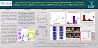
Home | Foundation | News & Media | Contact Us Perlecan – VEGF Signaling Perlecan Effects on Angiogenesis in a Perlecan-deficient Transgenic Mouse Hind-leg Ischemic ModelBeiping Qiang1,, Michael A. Kuliszewski2, Azriel Osherov1, Dmitriy Rudenko2,Paul Fefer1, Howard Leong-Poi2, Bradley H. Strauss1,1. Sunnybrook Health Sciences Centre, Schulich Heart Program, Toronto, Ontario, Canada; 2. Keenan Research Centre in The Li Ka Shing Knowledge Institute, St. Michael’s Hospital, Toronto, Ontario, Canada Saphenous artery Aorta Left External iliac artery Femoral artery Common iliac artery Abstract Introduction Methods Results Internal iliac artery Collaterals Background Atherosclerosis is the leading cause of coronary and peripheral arterial disease. Therapeutic angiogenesis could offer symptomatic relief for patients who are no longer candidates for conventional interventional therapies. Perlecan is a heparan-sulfate proteoglycan that is the major proteoglycan of basement membranes. Perlecan binds several angiogenic growth factors through the heparan moiety and modulates their activity. In this study, a hind-leg ischemic model was used to measure the influence of intact perlecan on angiogenesis in comparison to a perlecan-mutant (heparan deficient) transgenic mouse and determined its possible molecular mechanisms. Method We compared wild type and Hspg2Δ3/Δ3 mice (heparan-sulfate deficient perlecan transgenic strain) in a hind limb ischemic model of excision of the entire femoral arterial segment. A laser Doppler perfusion (LDPI) image analyzer (Moor Instruments) and Contrast-enhanced ultrasound (CEU) imaging of the hind limb abductor muscles were used to measure the ischemic/normal hind-limb blood perfusion ratio and to confirm ischemia and angiogenesis respectively. LDPI and CEU were performed at Day 2, Day 7, Day 14 and Day 28 after initial hind-leg ischemia surgery. At Day 28, after LDPI and CEU, the animals were euthanized and the hind-leg ischemic muscle harvested for histology, immunostaining , PCR and Western blotting to assess perlecan content and assays of VEGF and FGF, two heparin-binding angiogenic growth factors. ResultsLDPI showed significantly higher blood flow in ischemic limbs in wild-type compared to Hspg2Δ3/Δ3 mice at all time periods (Day 2: wild type 9.6±3.4%; mutant:6.9±1.6% P=0.038; Day 28: wild type: 61.3±16.3%; mutant: 41.1±11.7% P=0.002) . CEU showed normalized microvascular blood flow was significantly higher in the wild type compared with mutant at D28 (wild type: 0.667±0.12, mutant: 0.26±0.08 P=0.001). At Day 2, endogenous VEGF expression was significantly decreased in the ischemic limbs of Hspg2Δ3/Δ3 mice compared to wild type (P<0.05). Perlecan is a large (467-kDA) heparan sulfate proteoglycan (HSPG) that is expressed in most extra-cellular matrices (ECM) and basement membranes. It is the major extra-cellular HPSG in the blood vessel. Its core protein consists of five distinct domains which has exhibited one or more binding sites for a number of ligands, including basement membrane components, cell adhesion molecules , and growth factors. N-terminal domain I contains three attachment sites for heparan sulfate side chains, can function as a ligand for storage and release of heparan-binding growth factors (such as vascular endothelial growth factor [VEGF]), and also can protect these proteins from inactivation by proteolytic digestion. A potential pro-angiogenic mechanism of perlecan is VEGF binding to the HS side chain of perlecan, to enhance VEGF interaction with its receptor. To specifically address the role of HS side chains in perlecan in angiogenesis, we compared responses in a hind-leg ischemia model in Hspg2Δ3/Δ3 (HS deficient transgenic) and wild type (wt) mice. Popliteal artery Hind-leg Ischemia Model * Perlecan binds to VEGF and helps it migrate to its receptor Realtime reverse transcriptase-polymerase chain reaction data showing endogenous vascular endothelial growth factor (VEGF) is significantly upregulated in wildtype compared with Hspg2Δ3/Δ3 mice (P<0.05) • Hind Limb Ischemia Surgery • 66 mice (32mutant +34wildtype) Day 2 Day 28 Normalized microvascular blood flow by contrast-enhanced ultrasound in the ischemic leg was significantly higher in wild-type at D28 compared with perlecan-deficient mice (P=0.001) LDPI: n=49 (27 Hspg2Δ3/Δ3 ; 22 wt) @ D0, D2 , D7, D14 and D28 CEU:n=25 Wild type Right Leg Tail Left Leg D2: 8 Hspg2Δ3/Δ3 , 6 wt D28: 7 Hspg2Δ3/Δ3 , 4 wt Ischemia Normal Dissociation curve shows VEGF peak (left panel); Amplification plot shows the VEGF expression is markly higher in wildtype than in mutant mice (upper pannel) 40 samples for Realtime PCR, Western Blotting and Histology Normal Ischemia Mutant Left Leg Right Leg Tail Contrast enhanced ultrasound (CEU) perfusion images in wildtype and mutant mice 28 days after femoral artery ligation Conclusions Hspg2Δ3/Δ3 mice show a marked reduction in blood flow by both LDPI and CEU compared to wild-type mice in a hindlimb ischemia model. This is accompanied by decreased VEGF mRNA expression in Hspg2Δ3/Δ3 mice early after ischemia. Our data suggests that the HS side chains in perlecan are crucial for the angiogenic response, potentially through effects on VEGF mRNA expression. Further studies are planned to address this mechanism.


