Download
1 / 2
20 likes | 31 Views
This will be available on their web site when it is finished. limiting the quantity of radiation to which the person is revealed. In CT, the x-rays are targeted via the specific interior component of the body of interest at lots of numerous angles for every cross sectional slice.

E N D
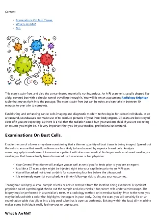
Content • • • Examinations On Bust Tissue. What Is An Mri? Mri. This scan is pain-free, and also the contaminated material is not hazardous. An MRI scanner is usually shaped like a big, covered box with a circular tunnel travelling through it. You will lie on an assessment Radiology Brighton table that moves right into the passage. The scan is pain-free but can be noisy and can take in between 10 minutes to over a hr to complete. Establishing and enhancing cancer cells imaging and diagnostic modern technologies for cancer individuals. In an ultrasound, soundwaves are made use of to produce pictures of your inner body organs. CT scans are best stayed clear of if you are expecting, as there is a risk that the radiation could hurt your unborn child. If you are expecting or assume you might be, it is very important that you let your medical professional understand. Examinations On Bust Cells. Enable the use of a lower x-ray dose considering that a thinner quantity of bust tissue is being imaged. Spread out the cells to ensure that small problems are less likely to be obscured by superior breast cells. Analysis mammography is made use of to examine a patient with abnormal medical findings-- such as a breast swelling or swellings-- that have actually been discovered by the woman or her physician. • • • • Your General Practitioner will analyze you as well as send you for tests prior to you see an expert. Just like a CT scan, a dye might be injected right into your capillaries prior to an MRI scan. You will be asked not to eat or drink for concerning four hrs before the ultrasound. It is extremely essential you schedule a timely follow-up visit to discuss your outcomes. Throughout a biopsy, a small sample of cells or cells is removed from the location being examined. A specialist physician called a pathologist checks out the sample and also checks it for cancer cells under a microscope. The biopsy may be performed in a specialist's areas, at a radiology method or in medical facility. Prior to the scan, you may be infused with a color that highlights the organs in your body. During the scan, you will certainly lie on an examination table that glides into a big steel tube that is open at both ends. Existing within the loud, slim machine makes some individuals really feel nervous or unpleasant. What Is An Mri?
2 radiology companies are advertising 'full body CT scans' of the whole body that will, they state, spot cancer prior to it's advanced and also comes to be tough to deal with. An ultrasound is a painless scan that makes use of soundwaves to create a picture of the within your body. The individual executing the ultrasound will spread out a gel on your skin, and afterwards move a tiny gadget called a transducer over the area. This sends out soundwaves that resemble when they fulfill something thick, like an organ or a tumor. a blood test to check for certain chemicals produced by cancer cells. This is a tool with a light and also a cam at one end as well as an eyepiece at the other. Many endoscopes have tiny devices that the medical professional can utilize to take biopsies of the body organs or cells being considered. Mri. You can figure out more regarding your danger of bust cancer by checking out the Family members background of bust cancer cells truth sheet. That is, they might provide a person a tidy costs of wellness, despite the fact that the person may in fact have a concealed cancer. That's because full body scans aren't as good at picking up cancers as more standard testing procedures - like colonoscopy to search for bowel cancers cells as an example. In some cases modifications will be seen on the ultrasound that were not noticed, or not believed to be very important, when the initial one was done. There is also study recommending that more surgical procedure is not constantly required for small areas of MRI-detected cancer. The added details from a bust MRI can help your specialist as well as oncology group plan your cancer treatment. In some cases a cancer that exists may not be located on the scan. Although it is an extremely sensitive test, MRI is not a perfect examination. It is still possible that an MRI scan will fall short to see a cancer cells in the bust due to the type and also/ or dimension of the cancer (a 'false adverse' outcome). Thumbs Down From Health Authorities. Although the variety of ladies as well as men being diagnosed is Australia is boosting, the number of deaths from bust cancer is lowering. Breast MRI is an exceptionally delicate, non-invasive, medical imaging evaluation utilized for detection of bust cancer cells and various other breast problems. It can additionally be used to take a look at the level of breast cancer cells after a medical diagnosis has been made adhering to a mammogram, ultrasound and/or biopsy. The high quality of photos created by MRI of bust tissue is superior. MRI makes it easy to take extra photos of the muscular tissue and chest wall around your breast to provide an extra extensive http://www.thefreedictionary.com/radiology diagnosis. MRI is able to develop extremely detailed images of the bust cells.