Download
1 / 20
200 likes | 346 Views
Nuclear pore complex proteins mark the implantation window in human endometrium. Elisa Guffanti1, Nupur Kittur1, Z. Nilly Brodt1, Alex J. Polotsky2, Satu M. Kuokkanen2, Debra S. Heller3, Steven L. Young4, Nanette Santoro2 and U. Thomas Meier1,*

E N D
Nuclear pore complex proteins mark the implantationwindow in human endometrium Elisa Guffanti1, Nupur Kittur1, Z. Nilly Brodt1, Alex J. Polotsky2, Satu M. Kuokkanen2, Debra S. Heller3, Steven L. Young4, Nanette Santoro2 and U. Thomas Meier1,* 1Department of Anatomy and Structural Biology and 2Department of Obstetrics, Gynecology and Womenʼs Health, Albert Einstein College of Medicine, 1300 Morris Park Avenue, Bronx, NY 10461, USA 3Department of Pathology, UMDNJ – New Jersey Medical School, Newark, NJ 07101, USA 4Department of Obstetrics and Gynecology, University of North Carolina School of Medicine, Chapel Hill, NC 27599, USA *Author for correspondence (e-mail: meier@aecom.yu.edu)
Summary • Nucleolarchannel systems (NCSs) are membranous organelle • appearing transiently in the epithelial cell nuclei of postovulatory human endometrium. • Their characterization and use as markers for a healthy receptive endometrium have been limited because they are only identifiable by electron microscopy. • Here we describe the light microscopic detection of NCSs using immunofluorescence
The monoclonal nuclear pore complex antibody 414 shows that NCSs are present in about half of all human endometrial epithelial cells but not in any other cell type, tissue or species. • Most nuclei contain only a single NCS of uniform 1 m diameter indicating a tightly controlled organelle. • The composition of NCSs is as unique as their structure; they contain only a subset each of the proteins of nuclear pore complexes, innernuclear membrane, nuclear lamina and endoplasmic reticulum.
Validation of our robust NCS detection method on 95 endometrial biopsies defines a 6-day window, days 19-24 (±1)of an idealized 28 day cycle, wherein NCSs occur. • Therefore,NCSs precede and overlap with the implantation window and serve as potential markers of uterine receptivity. • The immunodetection assay, combined with the hitherto underappreciated prevalence of NCSs, now enables simple screening and further molecular and functional dissection.
Introduction • Among the ultrastructuralhallmarksof secretory endometrial epithelial cells are giant mitochondria, subnuclear glycogen deposits, pinopodesand nucleolarchannel systems (NCSs) (Martel, 1981; Spornitz, 1992 • Whereas giant mitochondria and subnuclear glycogen deposits appear in the early luteal phase, pinopodes and NCSs more closely overlap with the mid-luteal window of implantation and could serve as potential markers (Clyman, 1963;Nikas et al., 1995).
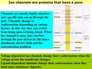
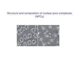
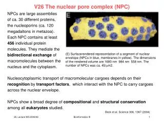
The NPCs are large protein assemblies consisting of 30 or so proteins (nucleoporins) present in multiple copies and arranged in partial symmetry across the envelope and around the pore. • NCSs have strictly been observed post ovulation, only on cycle days 16-24, and are not detected in pregnancy (Clyman, 1963). They appear to be induced by progesterone and are sensitive to oral and • intrauterine contraceptives
Finally, in several cases of unexplained infertility the absence or delayed appearance of NCSs was noted as the sole abnormal endometrial parameter (Dockery et al., 1996; Gore and Gordon, 1974; Kohorn et al., 1972). • Here we describe the first light microscopic detection and molecular dissection of NCSs.
Results Light microscopic detection of NCSs • we tested for the presence in NCSs of proteins from the nuclear boundary using indirect immunofluorescence on semi-thin frozen sections of human endometrium. • Indeed, the monoclonal antibody 414 (mAb414), directed against a subset of nuclear pore complex proteins (Davis and Blobel, 1986), identified rings in the nuclei of some endometrial epithelial cells
NCSs are abundant organelles specific to human endometrial epithelial cells • In single 0.5-μm-thick cryosections or 0.2-μm-thick optical confocal planes of paraffin sections, NCSs are observed in only about 10% of epithelial cell nuclei. • To assess the number of NCS in entire nuclei, 7-μm-thick paraffin sections were stained with mAb414 and imaged across their entire thickness in 0.2 μm steps using confocal laser-scanning microscopy. • NCSs were most abundant in epithelial glands but also present in luminal epithelium facing the uterine cavity. • on no occasion were NCSs observed in nuclei of stromal cells.
on no occasion were NCSs observed in nuclei of stromal cells. • analysis of tissue arrays containing six paraffin sections each of human esophagus, stomach, liver, colon, rectum, lung, kidney and breast tissue, failed to reveal any NCSs when stained with mAb414. • None of the carcinoma sections showed any. • Therefore, NCSs are restricted to the nuclei of healthy endometrial • epithelial cells.
The NCS marks the implantation window • Although previous electron microscopic studies agree that the NCS marks the postovulatory endometrium, the exact window of NCS appearance varies. • we tested our robust NCS detection method on 95 endometrial biopsies from fertile women: • 31 from the follicular phase • 64 from the luteal phase • NCSs were restricted to : • luteal days LH+4 to LH+13 • none were detected in any of the follicular phase biopsies • No NCSs were observed before day LH+4; after day LH+9 • the number of NCSs appeared to gradually decline and the number of cells with few and no NCSs increased.
Markers for uterine receptivity • Our identification of the first molecular markers of NCSs allowed development of a light microscopic assay for their detection. • Application of this assay reveals a peak presence of NCSs in over • 50% of endometrial epithelial cells or a tenfold higher prevalence • than was appreciated based on previous electron microscopic studies. • Therefore, our results establish the NCS as a major physiological hallmark of the postovulatory endometrium. • NCSs may be a hallmark of receptive endometrium because they define a luteal window that closely mirrors serum progesterone levels.
By contrast, pinopodes persist through early menses and pregnancy. • Here, we propose the long known, but mostly forgotten NCS as a marker for uterine receptivity.
Materials and Methods Human endometrial biopsies • Endometrial biopsies were obtained by informed consent from normally cycling women at two sites. • Endometrial tissue was fixed with 4% paraformaldehyde in phosphate-buffered saline. • Routine histological methods were used for paraffin embedding and sectioning of tissue. • Immunostaining of tissue sections
Fertil Steril. 2011 Mar 15;95(4):1385-9.e1. Epub 2010 Nov 10. The nucleolar channel system reliably marks the midluteal endometrium regardless of fertility status: a fresh look at an old organelle.