Download
1 / 1
20 likes | 148 Views
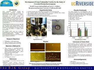
Pt Terminals. Digital Imaging Camera. Silicone Sealant. Inverted Microscope. Teflon Housing. Chamber designed by B2K. Electroformation Chamber. Function Generator. Oscilloscope. 50 microns. 25 microns. POPC GUVs. 25 microns. 25 microns. Egg PC GUVs. DPC GUVs.

E N D
Pt Terminals Digital Imaging Camera Silicone Sealant Inverted Microscope Teflon Housing Chamber designed by B2K Electroformation Chamber Function Generator Oscilloscope 50 microns 25 microns POPC GUVs 25 microns 25 microns Egg PC GUVs DPC GUVs Development of Giant Unilamellar Vesicles for the Study of Crowded Protein Environments David D. Gooray, Sandeep Dhall, and Victor G. J. Rodgers Bioengineering Department, University of California, Riverside Introduction Experimental Setup Results (Continued) Giant unilamellar vesicles (GUVs) are supramolecular structures consisting of amphiphiles that range in sizes from 10-100 µm.[1] They provide an easy method to: (1) observe environment for in-vitro studies of compartmentalized reactions and (2) model particular cell behavior. By studying the effects of crowded protein environments within GUVs, we will achieve a greater understanding of processes and properties such as mitochondrial swelling and osmotic pressure. Phosphatidylcholine lipids are the only lipids that can form GUVs under electroformation.[2] GUVs with a diameter ranging between 50-100 µm will withstand micromanipulation techniques and allow us to study the effects of crowded protein environments. We prepared GUVs using the 3 types of phosphatidylcholine lipids; 1-palmitoyl-2-oleoyl-sn-glycero-3-phosphocholine (POPC), L-α-phosphatidylcholine (Egg PC) and 1-2-dioleoyl-sn-glyeo-3-phosphocholine(DPC). • A square chamber was cut from teflon and molded to fit the microscope slide. • Two cylindrical platinum wires (Sigma-Aldrich) of 1.0 mm diameter were housed approximately parallel to each other with a 3.0 mm separation • Each wire was 1.5 mm from transparent viewing slide • All drilled holes and platinum-teflon interfaces were sealed with silicone • The GUVs selected for micromanipulation had an average size of 66.9 µm and were observed using a light microscope with a phase contrast filter. Average diameter of GUVs were determined using ImageJ. The average diameters of the GUVs obtained are: • POPC GUVs - 52.5 µm, • EPC GUVs - 67.6 µm, and • DOPC GUVs - 70.4 µm Research Objectives Experimental set up Size distribution of GUVs • Preparation of GUVs experiments • Obtain GUVs with 50-100 µm diameter • Develop a crowded protein environment Results Conclusion & Future Work We successfully setup the experiments for preparing GUVs and obtained the required sizes. GUVs with individual distribution on the wire were dependent on the precision of lipid application on the wire. The future direction includes loading of the GUVs with proteins while surrounding their environments with a similar concentration of the protein. This is to reduce the osmotic pressure within the GUVs and their respective loading characteristics. We will investigate better microinjection apparatus to enhance the precision of our piercings and load delivery into the GUVs. Materials & MethodsYa • Stock solutions of 1-palmitoyl-2-oleoyl-sn-glycero-3- phosphocholine (POPC), L-α-lysophosphatidylcholine (EPC) and 1-2-dioleoyl-sn-glyeo-3-phosphocholine (DPC) were made. (Avanti Polar Lipids) • Frequency: 10 Hz • A-C Voltage Ramp: 0.3 V increased to 2.3 V in 15 min • 2.3 V applied for 105 min Single GUV 25 microns References Acknowledgements [1]Luisi, P. and Walde, P. Giant Vesicles. New York: John Wiley & Sons Ltd. 2000 [2]Angelova,M.I., S.Soleau, P.Melead,J.- Faucon, and P.Bothorel. Preperation of giant vesicles by external ac electric fields: kinetics and applications.Prog. Colloid Polym. Sci. 89:127-131(1992) • B2K Group, • Dr. Vullev, Dr. Anvari, and Dr. Park • Jun Wang and Bioengineering department. GUVs that were obtained were found attached to the electrode. They will be micromanipulated for studying crowded protein environments.