Download
1 / 45
460 likes | 495 Views
Explore the antioxidant properties of polyphenolic compounds through complexing with metal ions, focusing on the chrysin-lanthanum complex. Learn about free radicals, antioxidants, and the importance of polyhydroxy phenols in biological systems.

E N D
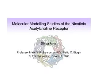
SYNTHESIS, SPECTROSCOPIC AND MOLECULAR MODELLING STUDIES ON LANTHANUM(III)-CHRYSIN COMPLEX
INTRODUCTION POLYHYDROXY PHENOLS Polyphenols are basically one of the most important oxygen donor compounds, which have been used as complexing agents for metal ions. These are characterized by the presence of multi hydroxyl groups in them, which offers a ready coordination site. Polyphenolic compounds found in fruits, vegetables and certain beverages that have diverse beneficial biochemical and antioxidant effects [1]. Antioxidant activity is due to metal ion chelation and scavenging active free radicals . They have been reported to have antiviral, antiallergic, anti-inflammatory, antitumor and antioxidant activities [2-4].
ANTIOXIDANTS Antioxidants are chemical substances that donate an electron to the free radical and convert it to a harmless molecule. Free radicals are highly reactive chemicals that attack molecules by capturing electrons and thus modifying chemical structures. Antioxidant play important role in biological system, such as suppressing the formation of active species by reducing hydrogen peroxide and by scavenging active free radicals [5]. What are free radicals? Any molecule or atom that possesses one unpaired electron is called “free radical”. These are unstable, short lived and will react with other compounds, which results in the stabilization of the free radical. Free radicals can be divided into different categories e.g., reactive oxygen species (ROS), reactive nitrogen species (RNS), reactive chlorine species (RCS) etc., The prominent member of such categories include superoxide (O2.-), hydroxyl (OH.), peroxyl (ROO.) and nitric oxide (NO.) radicals [6]
Generation of free radicals Free radicals generated in so many ways such as Fenton type reaction, where metal ions in biological system (eg. Fe3+/2+) involved and therefore a chain of oxidation and reduction reaction occurs which results in more free radicals [7]. Some of important sources of free radicals in biosystems are metabolism of drugs, inflammation, enzymatic activity, smoking and radiation [8,9]. Types of Antioxidant There are two mechanistic groups of antioxidants, those, which are interrupting the radical chain reaction, are called “chain breaking” (CB) antioxidants and those which inhibit or retard the formation of free radicals from their unstable precursors are called “preventive antioxidants”.
Naturally occurring antioxidants Both chain breaking and preventive antioxidants are naturally occurring in living bodies e.g., Vitamin E, Vitamin C, Polyphenols etc. Some important preventive antioxidants are catalase (CAT), glutathione peroxidase (GSHPX) and superoxide dismutase. FLAVONOIDS Flavonoids are polyphenolic compounds, which constitute one of the most characteristic classes of compounds in higher plants. The flavonoids have aroused considerable interest recently because of their potential beneficial effects on human health. They have been reported to have antitumor and antioxidant activities [2-4].
The chemical structure of flavonoid Antioxidant activity of flavonoids Flavonoids are powerful antioxidant, and their activity is related to their chemical structures. Plant polyphenols are multifunctional and can act as reducing agents, as hydrogen atom-donor and as singlet oxygen quencher.
AIM OF PRESENT WORK In present study, we have chosen chrysin (5,7-dihydroxy flavone) which shows interesting antioxidant activities and remarkable chelation with metal ions [10-13]. In order to investigate the interaction of chrysin with metal ion, spectroscopic (IR, UV-Visible, 1H NMR spectras and Thermal analysis) and molecular modeling studies were carried out. chrysin (5,7-dihydroxy flavone) • Biometals, 18; 143-154, 2005. • Bioorganic Chemistry, 33; 67-81, 2005.
MATERIALS AND THEORY OF METHODS USED Theory of Methods Used Although almost all parts of the electromagnetic spectrum are used for studying matter but energy absorption from four regions such as ultraviolet and visible, infrared, microwave and radio frequency absorption are of main concern. UV-Vis, IR and 1H NMR Spectroscopy Radiation absorbed Effect of the molecule UV-Visible Change in electronic energy (λ=200-800 nm) levels with in the molecules. Infrared Changes in vibrational and (=400-4000cm-1) rotational movements of the molecule Radio-frequency Changes in the magnetic (ν =60-600 MHz) properties of certain atomic nuclei (Hydrogen)
Thermogravimetric Analysis Thermal methods of investigation generally referred to as thermal or thermo- analytical techniques. These may be defined as experimental methods for characterizing a system (element, compound or mixture) by measuring changes in physico-chemical properties at elevated temperatures as a function of increasing temperature [14,15]. The two chief methods are • Differential thermal analysis (DTA) [16], in which changes in “heat content” are measured as a function of increasing temperature and • Thermogravimetric analysis (TGA), in which changes in weight are measured as a function of increasing temperature.
Computational Chemistry Computational chemistry is a new discipline. Its advent and popularity have paralleled improvements in computing power during the last several decades. As with other disciplines in chemistry, computational chemistry uses tools to understand chemical reactions and processes. Calculation Methods There are two types of methods in calculations:molecular mechanics and quantum mechanics. The quantum mechanics methods include semi-empirical, abinitio, and density functional quantum mechanics methods.
EXPERIMENTAL Materials Lanthanum oxide (Lieco Chemicals, USA) is converted to the corresponding chloride. Chrysin (Sigma-Aldrich, USA) and methanol (SD Fine) were used as such in this study. Preparation of La (chrysin)3 complex Hot solution of chrysin (3 m mol) in 50 ml methanol was added drop wise to the hot solution of hepta-hydrated lanthanum chloride (1m mol) in 50 ml methanol with constant stirring and kept the solution at 80oC temperature. After 1 hour stirring, yellow color needle shaped crystalline precipitate formed. The precipitate was filtered and washed with methanol and dried in vaccuo over P4 O10.
Methods or Physical Measurements Microanalysis (Carbon and Hydrogen) was carried out with a FI-SONS EA-1108 elemental analyzer. The metal content of the complex was estimated by complexometric titration. The thermogram was recorded on du Pont TA 2000 TGA machine under nitrogen atmosphere at a heating rate of 10oC min-1. Melting point (mp) was measured with a Gallen kamp MBF-595 apparatus. A Shimadzu UV-2501PC spectrophotometer was used to obtain the electronic spectra in the region 200-700 nm in methanol, DMF and DMSO solvents. FTIR spectra in the 4000-400 cm-1 regions were recorded from KBr pellets on a shimadzu-250 spectrophotometer. 1H NMR chemical shift was measured in DMSO-d6 solvent on Bruker 300 MHz spectrophotometer. Molecular modeling calculations were carried out using Hyperchem (Version 7.5)[17].
Results and Discussion The chrysin complex of lanthanum (III) has been isolated in methanol under normal conditions. The complex is pale yellow color polycrystalline solid and having sharp melting point between the range 289-290oC. The complex was hygroscopic in nature and soluble in common organic solvents but slightly soluble in water, ether, chloroform and CCl4. On the basis of elemental analysis, UV-Vis, IR, TGA/DTA and NMR spectral studies we assumed that chrysin acted as a bidentate ligand and formed a mononuclear complex where one La(III) ion is bound to three chrysin molecules. These results suggest that the composition of the complex is La(chrysin)3. Table 1. Physical properties of lanthanum-chrysin complex Complex Color MP oC % Metal Calculated (Observed) C H M La(chrysin)3 Yellow 289-90 51.84(50.96) 2.61(2.70) 13.32(12.72)
UV-VIS Spectral analysis The absorption spectra of the chrysin and its lanthanum complex were recorded in methanol, DMF and DMSO solvents within the spectral range 200-700 nm. The absorption spectra of free chrysin molecule revealed two major intense absorption bands in DMF, DMSO and methanol solutions in the ultraviolet part of the spectrum. The absorption in 260-326 nm range correspond to the B ring portion (phenolic system band I) and related to the П- П transitions with in the aromatic ring of the ligand molecules. The second absorption transition was observed between the spectral range 200-293 nm is correspond to the A ring portion (quinonolic system band II) which is due to П- П, n- П and n- transitions with in the quinonolic ring of the chrysin molecule.
The absorption spectrum of chrysin metal complex was shifted towards higher wavelength in comparison with free chrysin ligand spectrum. Such bathochromic shift can be explained by the extension of the conjugated system with the complexation. The absorption band II was shifted more than band I it indicates that band II (ring A, quinonolic system) is involved in coordination to the metal ion [18,19]. These observed results were suggested that 4-oxo and 5-OH groups of chrysin ligand are involve in coordination to the metal ion.
IR Spectra Infrared absorption spectra are found to be the most useful physical method for investigation & identifying functional groups. The characteristic IR absorption frequencies in the spectral range 4000-400 cm-1 were measured for free chrysin and its metal complex. The IR absorption spectrum of lanthanum metal complex clearly indicates that the free chrysin molecule loses their original characteristics and participate in coordination to the metal ion. The complex exhibited (M-O) band at 500-400 cm-1 while ligand exhibited no such band and suggests the metal-oxygen coordination. On the other hand, a diffused band in the region 3500-3000 cm-1 was appeared in the spectrum of ligand, which is attributed to the symmetrical and anti-symmetrical stretching modes of (O-H), which undergo change in the spectra of the complex. The appearance of this frequency suggests the presence of hydroxyl group and shift of the ligand frequency due to the loss of OH group during the coordination to the lanthanum ion.
The (C-O-C) and (C=C) frequencies changed slightly upon complexation indicates the ring oxygen does not form metal-oxygen bond. However, a strong band at about 1653 cm-1, detected in the spectra of the ligand is assigned to (C=O), which was shifted in the spectrum of the metal complex, which indicates that the coordination occurs through the C=O oxygen atom [20-22]. Table 3. IR absorption frequencies of lanthanum-chrysin complexe.
Thermal Analysis Thermogravimetric analysis of the complex was carried out to examine the thermal stability, number and nature of water molecule(s) present in the complex. The thermogram was recorded in the temperature range 30-600oC in nitrogen atmosphere with heating rate 10 oC per minute. Thermal spectra of the complex revealed that the complex is stable up to 300oC and does not show any weight loss below this temperature. This is strong evidence that the complex is devoid of lattice water as well as coordinated water in the coordination sphere. The TGA curve shows that the first weight loss of the complex was observed between the range 56.31-51.86 %, which occurs between the temperature 327-289 oC, corresponds to the two molecule of chrysin. After elimination of first and second molecule, decomposition of third molecule of chrysin was started simultaneously between the temperature 534-505 oC. The observed weight loss 28.15-27.23 %, which is equivalent to one molecule of chrysin.
Table 4. Thermal analysis data of lanthanum-chrysin complex.
1H NMR Spectra 1H NMR spectral studies have been carried out to investigate the solution structure of the lanthanum chrysin complex and its stability in the solution medium. The 1H NMR spectra of ligand and its lanthanum metal complex were measured in DMSO-d6 solvent. 1H NMR resonance signals with their tentative assignments are based on the reports available in the literature. The resonance signals of coordinated chrysin are found to have shifted downfield as well as up field as compared to the free chrysin molecule. The 5-OH proton resonance signal was not observed in the spectrum of complex. Disappearance of this resonance signal in the complex spectrum indicates that 5-OH proton loses during the complexation process and this phenolic group participates in coordination to the metal ion. These proton NMR spectrum results suggest that, DMSO is a strongly coordinating solvent, which coordinate to the metal ion and may replace the ligand from the coordination sphere and form the complex. The Uv-Vis spectral results also suggest that DMSO is strongly coordinating solvent. [23-28].
Table 5.1H NMR chemical shift data of lanthanum-chrysin complexe in DMSO-d6 on 300 MHz. S= singlet, d= doublet and m= multiplet
1H NMR Spectra of La(chrysin)3 complex in DMSO-d6 at 300 MHz.
Computational details Molecular modeling studies on chrysin and its complex were carried out using molecular mechanics (MM+) methods. The minimized energy of chrysin and La(chrysin)3 complex are 17.1360 and 70.7312 k.cal respectively. Geometry optimization of chrysin was also carried out via semiempirical (PM3) and ab initio methods to obtain charge density on atoms and geometrical parameters. Hydroxyl group (-OH) at 5th position and oxo group (-C=O) at 4th position have higher charge density; it suggests these two positions are most possible chelating sites.
Energy minimized structure of chrysin obtained by using semiemprical (PM3) and ab initio methods.
Energy minimized structure of chrysin and La(chrysin)3 complex obtained.[Cyanrepresents carbon and white andredrepresenthydrogen and oxygen]
charge density diagram of chrysin obtained by using semiemprical (PM3) and ab initio methods.
Conclusion UV-Visible, IR and 1H NMR studies on lanthanum complex and chrysin indicate that the metal ion coordinated through 5-(OH) and 4-(C=O) groups. Thermogravimetric study shows that the complex losses three molecules of chrysin. Molecular Modeling study also supports the above studies.
Reference • J. R. Corbett, The Biochemical Mode of Action of Pesticides. Academic Press, New York. 1974. • F. Matsumura, Toxicity of Insecticides. Plenum Press, New York. 1985. • R. D. O'Brien, Insecticides, Action and Metabolism. Academic Press, New York. 1967. • M. B. Shimkim, and N. N. Anderson, Proc. Soc. Exp. Biol. Med. 34(1936)135. • N. Noguchi, E. Niki, Radical Biolilgy & Medicine, 28(10)(2000)1538. • B. Halliwell, Biochemical pharmacol., 49(1995)1341 • H.J.H. Fenton, J. Chem. Soc.,65(1984)899. • J.J. Van Hammen, Nature(London), 231(1971)79. • P. Wardman, “Atmospheric oxidation and antioxidants” vol.III(Scott,G.Ed.), Elsevier, Amsterdam, 1993, chapter 4. • V. Cody, E. Middleton and J.B. Harborne, Plant Flovonoids in Biology and Medicine: Biochemical, Pharmacological and Structure Activity Relationships, Alan R.Liss, NY, (1986). • V. Cody, E. Middleton and J.B. Harborne and A. Beretz, Plant Flovonoids in Biology and Medicine II: Biochemical, Cellular and Medicinal properties, Alan R.Liss, NY, (1988). • J. Wilska-Jeszka, Wlad. Chem., 13(1959)289. • J. Pusz, B. Nitka and S. Wolowiec, Polish J. Chem., 75(2001)795.
J.P. Redfern, Editor,”Thermal Analysis Review”, Santon Instruments, London. • S. Gordon, “Encyclopedia of Sciences and Technology”, MecGraw-Hill Book Co.Inc., New York, Toronto and London, (1960)556. • R.C. Mackenzie and B.P. Mitchell, Analyst, 87(1962)420. • Hyperchem. Release 7.51 professional version, Hypercube Inc., Canada. • J. Kang, L. Zhou, X. Lu, H. Liu, M. Zhang and H. Wu, J. Inorg. Biochemistry, 98(2004) 79. • L.J. Porter and K. R. Markham, J. Chem. Soc.,(C), (1970)1309. • K. Nakamoto, Infrared and Raman Spectra of Inorganic and Coordination Compounds, 3rd Edit., John Wiley Interscience, New York, 1978, 264p. • J. R. Ferraro and W.R. Walker, Inorg. Chem. ,4(1965)1382. • V.T. Kasumov, E. Taş, F. Köksal and Ş. Ö.-Yaman, Polyhedron, 24(2005)319. • S. Quici, M. Cavazzini, G. Marzzani, G. Accorsi, N. Armaroli, B. Ventura and F. Barigelleti, Inorg. Chem., 44(2005) 529. • V. Gutmann, Coord.Chem. Rev., 18(1976) 225. • V. Monga, B.O. Patrick and C. Orving; Inorg. Chem., 44(2005) 2666. • U. Casellato, S. Tamburini, P. Tomasin, P.A. Vigato, S. Aime and M. Botta, Inorg.Chem.,38(1999)2906. • X.P. Yang, B.-S. Kang, W.K. Wong, C. Y. Su and H.Q. Liu, Inorg. Chem., 42(2003)169. • B. Bleaney, J. Mag. Resonance,8(1972) 91.