Download
1 / 21
280 likes | 560 Views
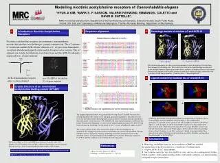
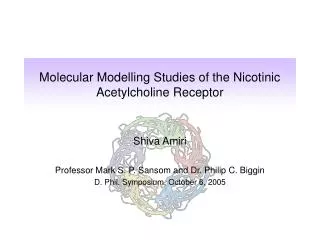
Molecular Modelling Studies of the Nicotinic Acetylcholine Receptor. Shiva Amiri Professor Mark S. P. Sansom and Dr. Philip C. Biggin D. Phil. Symposium, October 6, 2005. Ligand binding domain (LB) core of 10 β -strands, forming a β -sandwich

E N D
Molecular Modelling Studies of the Nicotinic Acetylcholine Receptor Shiva Amiri Professor Mark S. P. Sansom and Dr. Philip C. Biggin D. Phil. Symposium, October 6, 2005
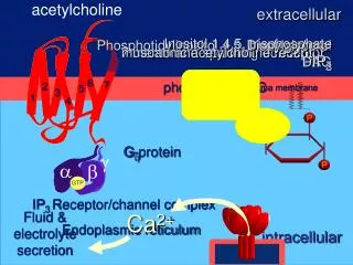
Ligand binding domain (LB) • core of 10 β-strands, forming a β-sandwich • an N-terminal α-helix, two short 310 helices • Transmembrane domain (TM) • 4 α-helices in each subunit (M1-M4) • Intracellular domain (IC) • α-helical, some residues still missing Unwin, Journal of Molecular Biology, March, 2005 The Nicotinic Acetylcholine Receptor (nAChR) • a ligand gated ion channel (LGIC) found in central and peripheral nervous system • endogenous ligand is acetylcholine (ACh) but reactive to many compounds such as nicotine, alcohol, and toxins • mutations lead to various diseases such as epilepsy, myasthenic syndromes, etc. • implicated in Alzheimer’s disease and Parkinson’s disease (not well understood) • mediates nicotine addiction
Computational methods to study membrane proteins • There are very few crystal structures available for membrane proteins • can build structures and use them to perform a range of studies such as electrostatics, pore profiling, ligand docking, Molecular Dynamics simulations etc. • Studying the movement of proteins to gain insight into their function • various methods of using a structure to look at the dynamics of the protein • Docking of ligands onto receptors • drug design
chosen {θ, z} theta (radians) z translation (Å) Scoring criteria x Possible gate region Generating Structures O. Beckstein, K. Tai Amiri et al., Mol. Mem. Biol, 2005
Atomistic Molecular Dynamics Simulations with GROMACS Coarse-grainGrouping of atoms Water Looking at the behaviour of water in the binding pocket Ligand Docking Docking Nicotine and other ligands onto various frames of trajectories In-house methodsGrouping data to simplify MD output CONCOORD Generating random structures from a given structure within distant constraints Gaussian Network Model (GNM)Assessing the flexibility of structures depending on the number of neighbouring residues Motion Analysis
ligand binding site Coarse-grain methods (1) • Gaussian Network Model (GNM) • A coarse-grained method to approximate fluctuations of residues based on the number of neighbours within a cut-off radius • Information on the flexibility of the receptor, may outline functionally important regions of the protein
SUB1 SUB2 SUB3 SUB4 SUB5 SUB1 SUB2 SUB3 SUB4 SUB5 Covariance matrix showing which part of the protein moves together. The red parts show highest covariance and the blue indicates negative covariance (move in opposite directions) Porcupine plot showing the nAChR’s two domains rotating in opposite directions. Suggests motions that could be involved in the gating mechanism http://s12-ap550.biop.ox.ac.uk:8078/dynamite_html/index.html (Barrett et al., 2004) Coarse-grain methods (2) CONCOORD • Generates n number of structures that meet distance constraints • very quick: one run takes a few minutes • Output used in Principle Component Analysis (PCA) to describe major modes of motion
Covariance lines show which sections of the receptor are moving together http://s12-ap550.biop.ox.ac.uk:8078/dynamite_html/index.html (Barrett et al., 2004) Molecular Dynamics Simulations • A method to study conformational changes at an atomistic level • MD of ligand binding domain of nAChR homologue, AChBP (Celie et al., 2004) • several simulations are being carried out for AChBP: • i) non-liganded simulations • ii) with various ligands: nicotine, carbamylcholine, HEPES • One nanosecond takes ~ 5 days for this system • Actual gating of this receptor happens on a millisecond time-scale
Molecular Dynamics Simulations continued … A simulation of AChBP with Nicotine in all 5 binding sites.
TRP 144 LEU 103 MET 115 THR 145 CYS 189 CYS 188 The important residues in the binding pocket are shown. These residues are thought to have key interactions with the ligand. Two subunits of the nAChR are shown with Nicotine inside the binding pocket The Binding Pocket • Studying structural changes which occur in the binding pocket to better understand how binding of a ligand results in the functioning of the ion channel • looking at distances, dihedrals of surrounding residues, and the behaviour of water in the binding pocket
Zone 2 Zone 1 Zone 4 Zone 5 Zone 3 Water in the Binding Pocket • Bridging waters form hydrogen bonds between the ligand and surrounding residues (shown using Ligplot) • Water seems to play an important role in ligand binding. There are various zones in the binding pocket where waters are frequently present
Water in the Binding Pocket Water molecules which remain in their position in the Binding Pocket
Docking • Docking various ligands such as nicotine, acetylcholine, imidacloprid (an insecticide) onto AChBP and the α7 nAChR to look at possible binding modes • An automated docking program docks ligands onto hundreds of frames from a trajectory Nicotine docked onto the AChBP binding site Nicotine Carbamylcholine HEPES
Results • Structure Generation: Developed a method to generate protein structures from their separate domains • Molecular Dynamics: • MD studies of AChBP with Nicotine, Carbamylcholine, and HEPES • Studying the role of water in the binding pocket • MD of α7 nAChR mutants • Coarse-graining: • GNM • CONCOORD • Grouping of information from MD trajectories • Automated Docking: Automated docking of ligands (Nicotine, acetylcholine, carbamylcholine, insecticides) onto AChBP and nAChR (and its mutants) along a trajectory
Prof. Mark S. P. Sansom Dr. Philip C. Biggin Dr. Alessandro Grottesi Dr. Kaihsu Tai Dr. Zara Sands Dr. Oliver Beckstein Dr. Daniele Bemporad Dr. Jorge Pikunic Dr. Andy Hung Dr. Shozeb Haider Dr. Syma Khalid Dr. Pete Bond Dr. Kia Balali-Mood Dr. Hiunji Kim Dr. Bing Wu Sundeep Deol Yalini Pathy Jonathan Cuthbertson Jennifer Johnston Katherine Cox Robert D’Rozario Jeff Campbell Loredana Vaccaro John Holyoake Tony Ivetac Samantha Kaye Sylvanna Ho Benjamin Hall Emi Psachoulia Chze Ling Wee Thanks to…
Future Work • Further investigation of the role of water in the binding pocket • Analysis of simulations of α7 nAChR mutants and docking along their trajectories • Development of further methods for understanding the motion of proteins from the limited structural data available • Combining coarse-grained and MD data…. i.e. Running GNM on various frames of a trajectory
Coarse-grain methods (3) Grouping eigenvectors • simplifying Molecular dynamics data by grouping the eigenvectors from the resulting trajectory.
TRP 144 LEU 103 MET 115 THR 145 CYS 189 CYS 188 The Binding Pocket • Studying structural changes which occur in the binding pocket to better understand how binding of a ligand results in the functioning of the ion channel • looking at distances, dihedrals of surrounding residues, and the behaviour of water in the binding pocket The important residues in the binding pocket are shown. These residues have been shown to interact with the ligand.
Using computational techniques to study the most flexible regions of the nAChR. These residues could play a key role in the gating of the receptor. Red shows the most flexibility, with blue showing least movement.