Download

1 / 51
510 likes | 673 Views
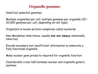
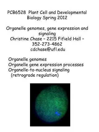
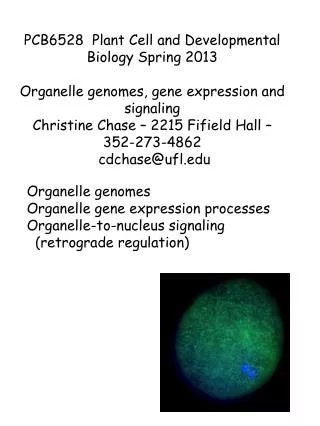
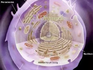
Organelle Presentations. Orange Block October 2012. The Nucleus. Emma Suneby and Elizabeth Young Orange Block. Structure. What does it look like? http://micro.magnet.fsu.edu/cells/nucleus/nucleus.html What is it made up of? Nuclear envelope, nucleolus, DNA, nucleoplasm. Location.

E N D
Organelle Presentations Orange Block October 2012
The Nucleus Emma Suneby and Elizabeth Young Orange Block
Structure • What does it look like? http://micro.magnet.fsu.edu/cells/nucleus/nucleus.html • What is it made up of? • Nuclear envelope, nucleolus, DNA, nucleoplasm
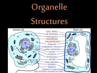
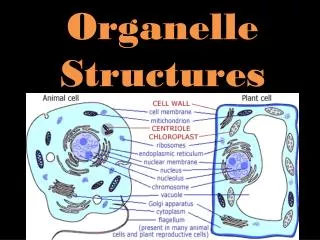
Location • Eukaryotic- inside both plant and animal cells, not located in bacteria • Located at the center of plant and animal cells • http://www.animalport.com/animal-cells.html • http://waynesword.palomar.edu/lmexer1a.htm
Function • Localizes the cells DNA • Tucks away DNA molecules making it easier for parent cells to copy genetic instructions before division • Outer membrane forms boundary in which cells control passages of substances and signals to and from the cytoplasm • Analogy: Nucleus is like the brain of the cell; it sends instructions for the rest of the cell. • Source: Biology Textbook The Unity and Diversity of Life
Nucleolus Chloé Kolbet and Molly Micou
Structure/Location • Inside the nucleus • Only in eukaryotic cells
Function • Where a large number of protein and RNA molecules (chromatin) are constructed • Houses the basic components of the cell • Chromatin are Subunits of ribosomes that go out of the nucleus’ pores to cytoplasm • Factory of sweaters • Source: Biology: The Unity and Diversity of Life by Starr and Taggart
Structure • Each ribosome organelle is made up of 2 subunits. • These are assembled inside the nucleolus, and the ribosome is therefore eukaryotic. • The ribosome can travel inside of cells if it is attached to the ER or float around the cytoplasm. • The ribosome is in animal, plant and bacteria cells.
Analogy • DNA is like an old book in a library that you can’t check out. • The mRNA is the copy of one specific recipe. For example, delicious chocolate chip cookies. • The ribosome is the kitchen where these cookies are made. • The mRNA or recipe goes to this place (ribosome or kitchen) and then attracts the specific enzymes or ingredients. • The end result is a chocolate chip cookie or a protein!
The two subunits (one large and one small) are like the top and bottom of a hamburger bun. They sandwich the mRNA (or hamburger).
Function • Polypeptide chains for proteins are assembled on the surface of the ribosome. • Ribosomes make these polypeptide chains by attracting specific enzymes. • The place where the mRNA (the instructions) binds to the surface.
Function: Part of Cytomembrane system; modifies polypeptide chains into final protein product and mRNA from nucleus Newly forming chains with specific strings of amino acids enter the space in the rough ER There, enzymes attach oligosaccharides and other side chains to the polypeptide, and modify the polypeptide within the ER into the final protein Sends out the new polypeptides in vesicles to the Golgi Body Structure: Arranged in stacks Made of up space(inside the ER) and ribosomes
http://www.tutorvista.com/content/biology/biology-iii/cell-organization/membranous-cell-organelles.phphttp://www.tutorvista.com/content/biology/biology-iii/cell-organization/membranous-cell-organelles.php
Location: • Only in eukaryotes • Surrounds the nucleus • It is in plant and animal cells. • Analogy: • Carpenter sanding and painting the final product.
http://sbaran.net/7science/cells/index.html http://www.animalport.com/animal-cells.html
by: Ashton Chryssicas and Ellen Kitsos Golgi Body
Structure/location • Layered, bean-like membranes • Similar appearance to a series of flattened pancakes • Eukaryotic cells • ribosomes are found in the pancake-like stacks. • Found in plant and animal cell • Floats in the cytoplasm
Function • Modifies polypeptide chains into mature proteins • Sorts, packages and distributes proteins and lipids (macromolecules) for secretion outside of cell • Absorbs vesicles from Rough ER • Vesicles form as patches of the membrane bulge out, then break away into cytoplasm. • Nutrients enter through the cell membrane and into the cytoplasm by Endocytosis • Vesicles gravitate towards cell membranes and are released in a process called exocytosis Analogy: The function of a golgi body is similar to that of a post office.
References • http://www.johnkyrk.com/golgiAlone.swf • Biology: The Unity and Diversity of Life • http://www.biology4kids.com/files/cell_golgi.html
Smooth E.R. (Endoplasmic Reticulum)
Smooth ER • FUNCTION: Lipid synthesis; Detoxifies certain compounds; In sarcoplasmic reticulum: located in skeletal muscle cells help in muscle contractions. • LOCATION: In Eukaryotes; Its in both plants and animal cells; It is a continuation of the rough ER; the difference is that it lacks ribosomes.
Structure It is Tubular It is made up of membranes.
Analogy • Its is a kitchen because thats where food is made and its sent out for people to eat it afterwards.
Chloroplasts Taylor Barnhill • Location: photosynthetic eukaryotic cells, all over the cell • Disk shaped, encased by two membranes • Function: Sunlight energy ATP • ATP = chemical energy, used to make sugars and organic compounds.
Thylakoid membrane has light-trapping pigments, chlorophyll, enzymes, and proteins which absorb energy and store it in the form of ATP energy • Grana = disks of thylakoid membrane (folded and stacked) • Stroma the semi fluid interior where ATP energy is used to make sugar, starch, and other organic molecules • Stromal lamellae = connect the grana
Analogy: A chloroplast is like a factory with an assembly line, each stage helps convert raw materials into useful products. • Sunlight ATP sugars, starch, organic materials. http://hyperphysics.phy-astr.gsu.edu/hbase/biology/chloroplast.html
Mitochondria By Peter and Liam
Structure • The outermost membrane faces the cytoplasm. • Inner membrane forms cristae, meaning it folds back on itself. • The space between both membranes are where hydrogen ions are stored • Resembles bacteria in biochemistry and size
Location & Function • Mitochondria exist only in eukaryotes, but are in both animal and plant cells. • There are at least 1 in each cell • They exist anywhere in the cell membrane. • Mitochondria contain their own DNA • They aid cell respiration
Analogy • Like the lungs of the cell • Help the cell to breath
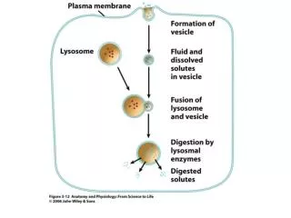
LYSOSOMES • FUNCTION • Used in intracellular digestion • Contain enzymes that speed the breakdown of proteins, complex carbohydrates, nucleic acids, and some lipids • Apoptosis - programmed cell death • Lysosomes release enzymes which break down cell components • Cancer cells cannot go through apoptosis, so they can’t kill themselves • LOCATION • In eukaryotic cells • All animals • Some plants • Some bacteria • In the cytoplasm of the cell
LYSOSOME STRUCTURE LYSOSOME IN A CELL • Bag-like structures • Different enzymes contained by a single membrane • This membrane protects from apoptosis happening when it shouldn’t
LYSOSOMES • http://www.ncbi.nlm.nih.gov/pubmed/18662570 • http://www.buzzle.com/articles/lysosome-structure.html • http://www.proprofs.com/flashcards/cardshowall.php?title=human-anatomyphysiology • http://www.biology4kids.com/files/cell_lysosome.html • Pencilcase • Case – membrane • Pens and pencils – enzymes • Pens can explode - apoptosisis
Function • Increases cell surface area • Storage for amino acids, sugars, ions (similar to a battery: stores energy and keeps everything running/moving) • Gets rid of toxic waste (face wash: gets rid of the bad parts of skin, keeps/ restores nutrients and hydration) • Causes fluid pressure in cell when enlarges
Structure • Made up of a fluid (cell “sap”) called tonoplast that is released into the cell when enlarges • Fluid provides nutrients for cell
Location • Eukaryotic • Located in the cytoplasm (narrow area between the central vacuole and the plasma membrane) http://www.cod.edu/people/faculty/fancher/cellstructure.htm • Can take up to 50%-90% of the cell interior • Found mainly in plant cells
Cell Membrane • Function: Controls the material exchanges and cell environment in interactions, an example of an exchanges is osmosis • Location: the outermost layer of the cell for animal cells, in plant and bacteria cells it is inside the cell wall • The cell membrane is found in all three types of organisms • Analogy: Security Guard
Composition of the Cell Membrane • Phospholipids Bi-layer • Proteins (ion channels and transmembrane protein channels) • Nuclear Pores
Function of Cell Wall The cell wall provides protection and structural support in plant and bacteria cells.
Structure of Cell Wall • Permeable to allow water and solutes to pass through • Middle Lamella-outermost layer, bonds with other cells • Primary Wall-made of gluey polysaccharides, glycoproteins, and cellulose (in plants) and peptidoglycan (in bacteria) which form into "rope-like strands" that are sticky, and cement cells together, it's thin and pliable and enlarges when water enters • Cuticle (a translucent, protective surface) forms when cells are exposed to air, keeps water from escaping • Secondary Wall- rigid to reinforce cell shape - in woody plants, made of lignin ( 3 carbon ring chain and an oxygen atom attach to 6 carbon ring structure)
Location of Cell Wall • Found in prokaryotes and some eukaryotes • Wrapped around the plasma membrane • Found in plant and bacterial cells, NOT animal cells
References Starr, Cecie, and Ralph Taggart. Biology: The Unity and Diversity of Life. 9th ed. USA: Thomson Learning, 2001. Print. http://images.tutorvista.com/content/cell-organization/cell-wall-layers.jpeg
Cytoskeleton By Ross Halpern & John Wilson
Cytoskeleton in Eukaryotic Cells The Structures and Functions of the Cytoskeleton The cytoskeleton is an organized network of two to three primary protein filaments: microtubules and micro/actin filaments which are present in Protista, Fungal, Plant, and Animal cells. Intermediate filaments are found in some animal cells only. The Cytoskeleton is a rope/ weblike structure found throughout the entirety of the cell that is responsible for nearly all Eukaryotic cell movement. Cytoskeleton of Animal Fibroblast cells. The microfilaments are tinted green while the microtubules are tinted orange.
Function of Cytoskeleton in Eukaryotic Cells • Establishes cell shape • Provides mechanical structure and internal organization • Intercellular transport of organelles • Gives cells capacity to move • Reinforces the plasma membrane Microtubules: Long, hollow cylinders made from monomers of the protein Tubulin. The largest part of the cytoskeletal system. They govern the division of cells as well as some aspects of their shape and many cell movements. Microfilaments: The thinnest of cytoskeletal elements. Made from 2 polypeptide chains of monomers from the protein Actin that are helically twisted together. Help with cell movements especially on the surface of the cell and the development of cellular shape in animal cells. Intermediate filaments: Most stable elements of cytoskeleton. Helps cells mechanically strengthen their parts and maintain their shape.