The Skeleton
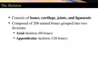
The Skeleton. Two Divisions. Axial Appendicular. Axial Skeleton. The “axis†of the Body. Skull Inner ear bones Hyoid Bone Rib cage Vertebral column. Axial Skeleton Functions. Framework for supporting and protecting organ systems in dorsal and ventral body cavities
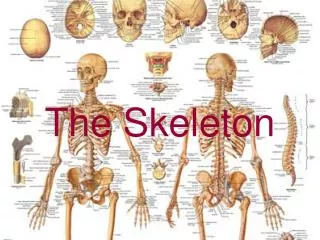

The Skeleton
E N D
Presentation Transcript
Two Divisions • Axial • Appendicular
The “axis” of the Body • Skull • Inner ear bones • Hyoid Bone • Rib cage • Vertebral column
Axial Skeleton Functions • Framework for supporting and protecting organ systems in dorsal and ventral body cavities • Surface area for muscle attachment • Head, neck and trunk stability and movement • Respiratory movement • Stabilize/position appendicular skeleton
Skull • Protect Brain • Support sense organs • Vision • Hearing • Balance • Olfaction • gustation
Skull • 22 bones • 8 cranial • 14 facial • Seven additional bones in the skull • 6 auditory ossicles • Hyoid bone
Hyoid Bone • Suspended below the skull by ligaments • Muscle base for the larynx (voice box) • Supports and positions the larynx
Vertebral Column • Spine is 26 bones • 24 vertebrae • Saccrum • Coccyx
Vertebral Column • Vertebrae are in regions • Cervical (C1 – C7): C1 = atlas; C2 = axis • Thoracic (T1 – T12) • Articulate with ribs • Lumbar (L1 – L5) • Total length in average adult is 28 inches
Intervertebral Disc • Fibrocartilage disc that lies between two adjoining vertebrae • Not found in sacrum or coccyx • “Shock absorbers”
Act as ligaments that hold the vertebrae of the spine together and as cartilaginous joints that allow for slight mobility in the spine. • Allow for movement at the waist as they act as a pivot point and allow the lumbar spine to bend, rotate, and twist
Vertebrae Anatomy • For the three types of vertebrae there are different distinguishing features • The openingsof the vertebrae(foramen) form thevertebral canalwhich enclosethe spinal cord
Vertebrae Anatomy • Vertebral foramen: opening • Vertebral arch: posterior margin of foramen • Transverse process: site for muscle attachment • Spinous process: Bump down your back • Body: weight-bearing portion • Lamina: roof of vertebral arch • Pedicle: walls of vertebral arch
Cervical Vertebrae • There are seven cervical vertebrae which are located in the neck. • They are the smallest, and lightest vertebrae of the vertebral column.
Cervical Vertebrae Anatomy Spinous Process Superior articular facet Lamina Foramen Pedicle Transverse Process Body
Thoracic Vertebrae • The rib cage of the chest is attached to the thoracic spine at each level. • Gives a great deal of stability and support to the upper body. • Limits the back's movement at the chest level.
Thoracic Vertebrae Anatomy Spinous Process Transverse Process Lamina Superior articular facet Foramen Pedicle Body
Lumber Vertebrae • There are 5 lumbar vertebrae located in the lower back. • Receive the most stress and are the weight-bearing portion of the back. • Allow movements such as flexion and extensionand some lateral flexion.
Lumbar Vertebrae Anatomy Spinous Process Superior articular facet Lamina Foramen Pedicle Transverse Process Body
Sacrum and Coccyx • Sacrum: five fused vertebrae • Protects reproductive and digestive organs • Attaches axial to appendicular skeleton • Extensive muscle attachment • Coccyx: 3-5 fused vertebrae • Attachment site for muscle that closes anal opening
Spinal Curves • Curved to allow for weight distribution • 2 primary curves: appear in late fetal development • Thoracic • Sacral • 2 secondary curves: occur months after birth • Cervical • lumbar
Spinal Curves Secondary Curve Primary Curve Primary Curve Secondary Curve
Chest Bones (Thorax) • Thoracic Vertebrae • Ribs • Sternum
Ribs and Sternum • 12 pairs of ribs • 7 pairs of “true ribs” • Reach the anterior body wall and connect to the sternum by separate cartilage (costal cartilage) • 8-12 are “false ribs” • Do not attach directly to the sternum • Costal cartilage of 8-10 fuses with 7 • Last Two pairs = “floating ribs” • No sternum connection
Sternum • Manubrium: articulateswith the clavicle • Body • Xiphoid process
intervertebral disc x ray • http://www.chirogeek.com/000_disc_anatomy.htm • http://spanky.thehawkeye.com/features/surgery/index.html