Download
1 / 39
410 likes | 559 Views
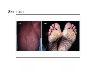
A 3 year old girl presents with high fever,extensive skin rash,and conjuctival congestion. Physical examination reveals cervical lymphadenopathy,erythematous palms and soles,and dry and red oral mucosa.

E N D
A 3 year old girl presents with high fever,extensive skin rash,and conjuctival congestion. Physical examination reveals cervical lymphadenopathy,erythematous palms and soles,and dry and red oral mucosa.
A throat culture is negative. Routine CBC is normal. The monospot test for infectious mononucleosis is negative. Additional tests for CMV and toxoplasmosis are negative as well.
Two months later she develops signs and symptoms of heart failure and goes into cardiac arrest. What is the appropriate diagnosis?
Vasculitis • Inflammation of the vessel wall. • Signs and symptoms: 1- local findings according to the involved tissue 2- systemic manifestations (fever, myalgia, arthralgias, and malaise)
vasculitis can be classified according to vessel size, immune complexes, specific autoantibodies, granuloma formation, etc • considerable clinical and pathologic overlap among many of these disorders can be present
Pathogenesis 1- Immune-mediated inflammation 2- Direct vascular invasion by infectious pathogens 3- Physical injury (radiation, mechanical trauma) 4- Chemical injury (toxins)
Non-infectious vasculitis • The main immunologic mechanismsunderlying noninfectious vasculitis are: - Immune complex deposition - Antineutrophil cytoplasmic antibodies (ANCA) - Anti-endothelial cell antibodies - Autoreactive T cells
Immune complex deposition • is implicated in the following vasculitides: 1- Drug hypersensitivity vasculitis. • e.g., penicillin • vary from mild and self-limiting, to severe and even fatal • skin lesions are most common. • Treatment: discontinuation of the offending drug. 2- Vasculitis secondary to infections. • Antibody to microbial molecules form immune complexes • e.g 30% of patients with PAN immune complexes composed of (HBsAg) and anti-HBsAg antibody
Anti-NeutrophilCytoplasmic Antibodies • ANCAs = circulating antibodies that react with neutrophil cytoplasmic antigens (mainly enzymes) • ANCAs blood levels are very useful markers for diagnosis, clinical severity, and as predictive of disease recurrence.
Two types are most important: 1-Antiproteinase-3 (PR3-ANCA)= c-ANCA. is a neutrophil azurophilic granule constituent similar to numerous microbial peptides; e.g. Wegener granulomatosis. 2-Anti-myeloperoxidase (MPO-ANCA)= p-ANCA. MPO is a lysosomal granule constituent; e.g. microscopic polyangiitis and Churg-Strauss syndrome MPO-ANCAs are also induced by several drugs, e.g. propylthiouracil.
Anti-Endothelial Cell Antibodies • Antibodies to endothelial cells • Associated with certain vasculitides, such as Kawasaki disease
Giant Cell (Temporal) Arteritis • The most common form of vasculitis among the elderly in developed countries. • Chronic, granulomatous, inflammation of large arteries, mainly the temporal arteries. • Vertebral and ophthalmic arteries, as well as the aorta (giant cell aortitis), also can be involved. • Ophthalmic artery involvement sudden and permanent blindness (rapid diagnosis and treatment are mandatory)
Giant Cell (Temporal) Arteritis • Pathogenesis: T cell-mediated immune response to unknown vessel wall antigen. • Morphology: • changes are patchy along the length of affected vessels. • nodular intimal thickening & thromboses reduce the lumen diameter and cause distal ischemia. • granulomatous inflammation (75%) within the inner media centered on the internal elastic membrane (( lymphocytes and macrophages, with multinucleate giant cells )) • fragmentation of the internal elastic lamina. • lesions at different stages of development are seen within the same artery
Giant Cell (Temporal) Arteritis- clinical picture • rare before the age of 50. • Signs and symptoms: • fever, fatigue, weight loss • facial pain or headache, (superficial temporal artery). • Ocular symptoms (ophthalmic artery) in 50% of patients; range from diplopia complete vision loss. • Diagnosis: - biopsy and histology • Treatment: - Corticosteroid or anti-TNF therapies
Takayasuarteritis • a granulomatous vasculitis of medium-sized and large arteries • ocular disturbances + marked weakening of the pulses in the upper extremities ( the pulseless disease). • scarring and thickening of the aorta. • the aortic arch with severe luminal narrowing of the major branch vessels. • the distinction from giant cell aortitis is made largely on the basis of a patient's age: >50 years giant cell aortitis <50 yearsTakayasu aortitis. • Pathogenesis: An autoimmune etiology is likely
Takayasu arteritis • Affects the aortic arch and arch vessels (2/3) • Pulmonary arteries (50% of patients) • Renal and coronary arteries also can be affected.
The histologic picture: a spectrum ranging from: • Mild WBC infiltrates • Intense transmural inflammation • Granulomatousinflammation, with giantcells and patchy medial necrosis • Irregular thickening of the vessel wall • Intimal hyperplasia • Adventitial fibrosis.
Clinical Features of Takayasu Aortitis • Initially, nonspecific: fatigue, weight loss, and fever. • With progression: • Reduced upper limb B.P. and pulse; neurologic deficits • Ocular: visual defects, hemorrhages, blindness. • Leg claudication distal aorta involvement • Pulmonary hypertension pulmonary artery involvement • MI Narrowing of the coronary ostia • Hypertension renal arteries (50% of patients).
Disease evolution: • rapidly progressive in some cases • quiescent after 1 to 2 years (visual or neurologic deficits) in others
Polyarteritis nodosa (PAN) • Systemic vasculitis of small or medium-sized muscular arteries • Typically involves the renal and visceral vessels and spares the pulmonary circulation. • There is no association with ANCAs - (1/3) chronic hepatitis B infection immune complexes containing hepatitis B antigens deposit in affected vessels. - (2/3) The cause is unknown
Polyarteritis nodosa (PAN)- morphology • Kidney , heart, liver, and GIT vessels are affected in descending order of frequency. • Segmental transmural necrotizing inflammation of small to medium-sized arteries, often with superimposed thrombosis. • ulcerations, infarcts, ischemic atrophy, or hemorrhages in the affected tissues • Inflammation weakens arterial wall aneurysms and rupture • Lesions have different stages at the same vessel and at any given time
PAN- The clinical course • disease of young adults but can occur in all age groups. • episodic, with long symptom-free intervals. • malaise, fever, and weight loss • the vascular involvement is widely scattered. • A "classic" presentation can be: a combination of: - malignant hypertension (renal artery) a major cause of death - abdominal pain and bloody stools (GIT lesions) - muscular aches and pains - peripheral neuritis. • Treatment: untreated fatal - immunosuppressionremission or cure in 90% of the cases
Kawasaki disease • acute, febrile, usually self-limited illness of infancy and childhood (80% of cases < 4 years) • arteritis of mainly large to medium-sized vessels. • Involvement of coronaryarteries aneurysms rupture or thrombosis myocardial infarction. • Originally in Japan, the disease is now recognized worldwide • Pathogenesis: anti-endothelial cell antibodies • Treatment: • intravenous immunoglobulin therapy and aspirin • Nowadays, symptomatic coronary artery disease < 4%.
Kawasaki disease • Acute phase=dense transmural inflammatory infiltrate • Chronic phase= aneurysm formation, intimal thickening. • Mucocutaneous lymph node syndrome: • conjunctival and oral erythema and blistering • erythema of the palms and soles • a desquamative rash • cervical lymph node enlargement.
Complications: • 1-cardiovascular disease (20% of untreated patients): ranging from asymptomatic coronary arteritis coronary artery aneurysms (7 to 8 mm) with rupture or thrombosis • 2-MI • 3-Sudden death.
Microscopic Polyangiitis • Hypersensitivity vasculitis or leukocytoclasticvasculitis • Necrotizing vasculitis affects capillaries, smallarterioles and venules. • All lesions tend to be of the same age in any given patient. • The skin, mucous membranes, lungs, brain, heart, GIT, kidneys, and muscle • Necrotizing glomerulonephritis (90% of patients) • Pulmonary involvement is common. • Pathogenesis: unknown
Microscopic polyangiitis- morphology • segmental fibrinoid necrosis of the media with focal transmural necrotizing lesions • granulomatous inflammation is absent. • infiltrating neutrophils frequently undergo fragmentation, hence the term leukocytoclastic vasculitis. • most lesions are "pauci-immune" (i.e., show little or no antibody or immune complexes)
Clinical Features of Microscopic Polyangiitis • Depending on the vascular bed involved: • Hemoptysislung • Hematuria, proteinuria kidney • Abdominal pain or bleedingGIT • Muscle pain or weakness muscles • Palpable cutaneous purpura skin • Treatment: - immunosuppression -removal of the offending agent
Wegener granulomatosis • is a necrotizing vasculitis • specific triad of: 1- Granulomas of the lung and/or the upper respiratory tract (ear, nose, sinuses, throat) 2- Vasculitis of small to medium-sized vessels (capillaries, venules, arterioles, and arteries) mostly in lungs and upper respiratory tract 3- Glomerulonephritis: (FSGS) (crescentic glomerulonephritis)
Wegener granulomatosis- pathogenesis • cell-mediated hypersensitivity response against inhaled infectious or environmental antigens. • PR3-ANCAs (c-ANCA) > 95% of cases - are useful markers of disease activity (After immunosuppressive therapy, ANCA levels fall dramatically, while rising titers are predictive of relapse) • The typical patient is >40 year old and male, although women and all ages can be affected. • If untreated, the mortality rate at 1 year is 80%.
Wegener granulomatosis- clinical picture • rash, myalgias, articular involvement, neuritis, and fever • bilateral pneumonitis, nodules and cavitary lesions (95%) • chronic sinusitis (90%) • mucosal ulcerations of nasopharynx (75%) • renal disease (80%)hematuria, proteinuria rapidly progressive renal failure. • Treatment: steroids, cyclophosphamide, TNF inhibitors and anti-B cell antibodies • Most patients with Wegener granulomatosis now survive, but remain at high risk for relapses that can ultimately lead to renal failure.
Churg-Strauss syndrome • allergic granulomatosis and angiitis • is a small vessel necrotizing vasculitis • classically associated with asthma, allergic rhinitis, lung infiltrates, peripheral eosinophilia, necrotizing granulomas, infiltration by eosinophils. • extremely rare disorder. • Pathogenesis: Unknown
Purpura, GIT bleeding, and renal disease (FSGS) are the major associations. • Cardiomyopathy: due to eosinophils (in 60% of patients) a major cause of morbidity and death.
Thromboangiitis obliterans (Buerger disease) • a disorder of severe vascular insufficiency and gangrene of the extremities. • focal acute and chronic inflammation of medium-sized and small arteries, especially the tibial and radial arteries, associated with thrombosis • secondary extension into adjacent veins and nerves may be seen.
Pathogenesis: • almost exclusively in heavy tobacco smokers and usually < age 35. • The etiology is unknown: components of tobacco- ? Direct endothelial cell toxicity ? an immune response. ? A genetic predilection increased prevalence in certain ethnic groups
Thromboangiitisobliterans (Buerger disease)–clinical manifestations • Early : Raynaud phenomenon, foot pain induced by exercise, superficial nodular phlebitis (venous inflammation). • severe pain-even at rest neural involvement. • Chronic ulcerations • Gangrene of fingers and toes • Treatment: Smoking abstinence in the early stages of the disease