Download
1 / 37
370 likes | 817 Views
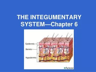
Alfred Ammoury Division of Dermatology, St George Hospital UMC. Embryology of the Integumentary system “The making of “. Layers of skin. Skin is a complex organ Horizontally stratified into 3 compartments -epidermis -dermis -subcutis (hypodermis) Vertically penetrated by appendages

E N D
Alfred Ammoury Division of Dermatology, St George Hospital UMC Embryology of the Integumentary system“The making of “
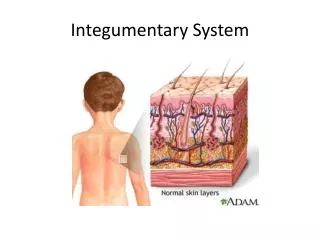
Layers of skin • Skin is a complex organ • Horizontally stratified into 3 compartments -epidermis -dermis -subcutis (hypodermis) • Vertically penetrated by appendages -hair follicles -sebaceous glands -eccrine and apocrine sweat glands
Week 2 – The Two-Layered Embryo • Bilaminar embryonic disc : Epiblast and the hypoblast
Week 3 – The Three-Layered Embryo • Primitivestreak – raised groove on the dorsal surface of the epiblast • Gastrulation – a process of invagination of epiblast cells • Endoderm – formed from migrating cells that replace the hypoblast • Mesoderm – formed between epiblast and endoderm • Ectoderm – formed from epiblast cells that stay on dorsal surface
Overview • Skin layers are derived from 2 different germ layers: • Ectoderm (lateral to the neural plate): epidermis • Mesoderm: dermis
Overview • Earliest skin study (4 week): single layered epidermis and a thin mesenchymal dermis. • Progressive development over the first 6 months: by the end of the second trimester, the skin is a stratified squamous epithelium. • Dermis lags behind epidermis in developemnt. • Dermal bulk increases post nataly and its maturation continues
Overview • Day 20-30: skin of embryo starts to develop (organogenesis). • Day 60: most organ systems have formed (including the skin). Embryo enters fetal period of growth and differentiation.
Epidermal development • Gastrulation occurs during the third week after fertilization • Complex process of involution and cell redistribution resulting in the formation of the three primary embryonal germ layers: Ectoderm, Mesoderm, endoderm
Epidermal development • Shortly after gastrulation, the ectoderm further subdivides into neuroectoderm and the presumptive epidermis that covers the embryo
At this stage, the ectoderm (presumptive epidermis) that covers the embryo is made of basal cells and superficial periderm cells
Periderm cells • Form a pavement epithelium that blanket the developing epidermis. • Exfoliated peridermal cells and sebum form the Vernix caseosa , a white greasy substance that covers the fetal body, protects it from amniotic fluid contents and facilitates delivery.
By the end of 8th week of gestation • Hematopoiesis has switched from the extraembryonic yolk sac to the bone marrow. The classical division between embryonal and fetal development. • Epidermal stratification occurs: formation of an intermediate layer, highly proliferative cells that eventually evolve into a multi layer structure that will replace the periderm.
Fetal Dermo-epidermal Junction • Basal layer begins to elaborate proteins that will anchor them to the developing basal lamina. • Late fetal development reveals the further differenciation of keratinocytes in the epidermis: granular layer, stratum corneum that replaces the periderm at W21. • Stratum germninativum starts to extend downward growth (ridges) into the developing dermis.
Specialized cells in the epidermis • Melanocytes, Merkel cells and Langerhans cells can be detected by the end of the embryonic period.
The melanocyte • Neural crest derived • W8 neural crest cells mgrates into mesenchymal tissue and differentiate into melanoblasts. • Melanoblasts localize to DEJ and hair bulbs, and differenciates into melanocytes
Langerhan cell • Bone marrow derived • Detectable by 40 days • By the third trmester most of the adult number of these cells will have been produced
Merkel cells • Slowly adapting touch receptors. • Controversial origin: neural crest Vs in situ differentiation of ectodermal cells. • Present at 11-12 weeks.
Dermis • Develops from mesenchyme • W11: mesenchymal cells produce collagenous and elastic tissue. • Part of the dermis project into the epidermis forming pappillae. • Capillary loops and nerves develop in these papillae • Major vasculature : by the end of the 1st trimester.
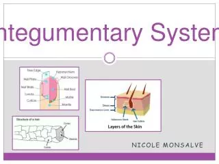
Appendegeal development • Hair follicles, sebaceous glands and sweat glands develop from the epidermis and they grow into the dermis.
Hair follicle • W12: Hair first appears on eyebrow & upper lip • 1st hair is lanugo hair (soft, fine, lightly pigmented and non mediullated). • Replaced by coarse terminal hair
Hair follicle • Stratum germinativum proliferates into the dermis. • Hair bud becomes club shaped=Hair bulb containing germinal matrix cells that willlatinize to form the hair shaft. • Hair bulbs are soon invaginated by small mesenchymal hair papilla. • Arrector pilorum develop from the surrounding mesenchyme.
Sebaceous glands • Buds from the sides of developing epithelial root sheath of hair follicles • Inactive until puberty.
Eccrine glands They develop as epidermal downgrowth Into the underlying dermis Starts at about W20 Functional at birth
Apocrine glands • Develop from downgrowths of the stratum germinativum that gives rise to hair follicle • Begin secretion around 7-10 years.
Nails • Fingernails reach the edge at W32 • Toenails reach the edge at W36
Mammary Glands • Highly modified and highly specialized type of sweat glands (development similar to that of SG) • W6: downgrowths of epidermal ridges in the pectoral area • Primary duct forms many 2ndary ducts that in turn develop to form lactiferous duct (15-20 at term). • Epidermis at the site forms a shallow pit mammary pit where the nipple will develop.