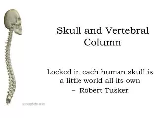
VERTEBRAL COLUMN
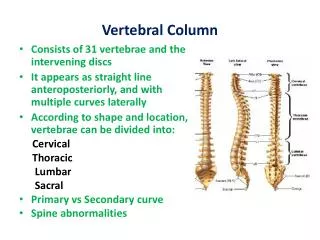
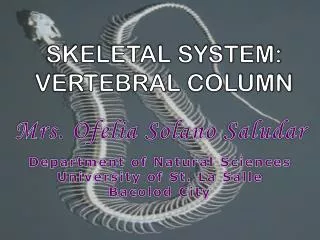
VERTEBRAL COLUMN. Gross Anatomy. 28 inches in length Medial border of scapula: Runs parallel to vertebral column Crosses ribs 2-7. Vertebral Column Regions. Cervical (7) Thoracic (12) Lumbar (5) Sacral (5) Coccygeal (3-4). Primary Normal Curvatures. Present at birth


VERTEBRAL COLUMN
E N D
Presentation Transcript
Gross Anatomy • 28 inches in length • Medial border of scapula: Runs parallel to vertebral column Crosses ribs 2-7
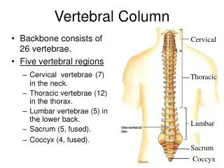
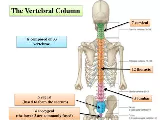
Vertebral Column Regions • Cervical (7) • Thoracic (12) • Lumbar (5) • Sacral (5) • Coccygeal (3-4)
Primary Normal Curvatures • Present at birth • Convex posteriorly • Examples: Thoracic Sacral • Exaggeration: Kyphosis
Secondary Normal Curvatures • Acquired later • Concave posteriorly • Examples: Cervical Lumbar • Exaggeration: Lordosis
Curvatures • Scoliosis • Spondylosis: Refer to text: p 496
Intervertebral Articulations • Disc (symphysis) • Synovial • Sacroiliac: Part synovial and part fibrous • Costal facets and demifacets
Disc (symphysis) • 20-30% of length of the column • Thicker in cervical and lumbar regions: Greater flexibility • Thinner in thoracic region: Reduced flexibility • Structure: Annulus fibrosus (fibrocartilage) Nucleus pulposus (avascular)
Synovial Joints • Plane joints • No joint capsules • Between superior and inferior articulating facets: Direction of motion determined by orientation of facets
Motion Segment • Functional unit of the spine • Consists of: Two vertebrae Intervening soft tissue Anterior segment: Two bodies, disc, longitudinal ligaments. Posterior segment: Two neural arches, apophyseal joints, intervening ligaments.
Cervical Vertebrae • Transverse foraminae: For vertebral arteries • Oblique articular facets • C2-C6 spines usually bifid • C7 = vertebra prominens • C1 = atlas • C2 = axis
Thoracic Vertebra • Have ribs attached: Bicipital ribs: To centrum To transverse process • Centrum is heart-shaped • Articular facets more horizontal • Spinous processes longer and narrower
Typical Thoracic Vertebra • Has two costal demifacets on body: Superior: For articulation with head of own rib. Inferior: For articulation with head of inferior rib. • Has costal facets for tubercle of rib on transverse process for own rib.
Atypical Thoracic Vertebrae • #1: Complete costal for own rib. Inferior costal demifacet on body for rib 2. • #10: Complete costal facet on pedicel and body. May have costal facet on transverse process. • #11, 12: Costal facet on each pedicle. No costal facets on transverse processes.
Lumbar Vertebra • Large, bulky centrum • Broad spinous processes • No rib articulations • Articular facets are sagittal • Mamillary processes: On back rim of each superior articular process. Origins for multifidi.
Sacrum • Consists of 5 fused vertebrae • Wing-like processes of each vertebra= Alae For attachment to ilium • Lip of body of first sacral vertebra = Sacral promontory
Sacrum • Posterior crests: Median crest: Fused spinous processes Intermediate crests: Fused articular (zygopophyseal) processes Lateral crests: Fused transverse processes • Sacral hiatus