The Vertebral Column
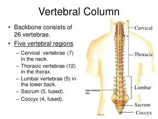
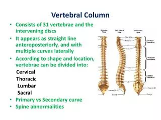
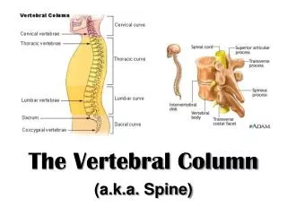
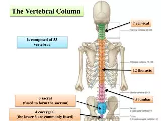
The Vertebral Column. 7 cervical. Is composed of 33 vertebrae. 12 thoracic. 5 sacral (fused to form the sacrum). 5 lumbar. 4 coccygeal (the lower 3 are commonly fused). A typical vertebra consists of : 1-a rounded body anteriorly 2-a vertebral arch posteriorly.

The Vertebral Column
E N D
Presentation Transcript
The Vertebral Column 7 cervical Is composed of 33 vertebrae 12 thoracic 5 sacral (fused to form the sacrum) 5 lumbar 4 coccygeal (the lower 3 are commonly fused)
A typical vertebra consists of: 1-a rounded body anteriorly 2-a vertebral arch posteriorly. They enclose a space called The vertebral foramen through which run the spinal cord and its coverings The vertebral arch gives rise to seven processes: a-One spinous b-Two transverse c- Four articular • The spinous process is directed posteriorly • from the junction of the two laminae. • The transverse processes are directed laterally • from the junction of the laminae and the pedicles • The articular processes are vertically arranged and consist of: • Two superior & Two inferior processes • They arise from the junction of the laminae and the pedicles • .
The pedicles • are notched on their • upper and lower borders • Forming • the superior and inferior • vertebral notches. On each side the superior notch of one vertebra and the inferior notch of an adjacent vertebra together form an intervertebral foramen. These foramina, in an articulated skeleton, serve to transmit the spinal nerves and blood vessels.
Characteristics of a Typical Cervical Vertebra • The transverse processes possess • a foramen transversarium • for the passage of the vertebral artery and veins • (note that the vertebral artery passes through the transverse processes C1 to 6 and not through C7). • The spines are small and bifid • The vertebral foramen is large and triangular
The first, second, and seventh cervical vertebrae are atypical. • The first cervical vertebra • THE ATLAS • does not possess a body or a spinous process • It has an anterior and posterior arch • It has a lateral mass on each side with articular surfaces on its upper surface for articulation with the occipital condyles • (atlanto-occipital joints) • and articular surfaces on its lower surface • for articulation with the axis • (atlantoaxial joints) Characteristics of the Atypical Cervical Vertebrae
The second cervical vertebra The AXIS has a odontoid process that projects from the superior surface of the body (representing the body of the atlas that has fused with the body of the axis). The seventh cervical vertebra or vertebra prominens is so named because it has the longest spinous process, and the process is not bifid. The transverse process is large, but the foramen transversarium is small and transmits the vertebral vein
Characteristics of a Typical Thoracic Vertebra • The body is heart shaped • The vertebral foramen is small and circular • The spines are long and inclined downward • Costal facets are present on the sides of the bodies for articulation with the heads of the ribs • Costal facets are present on the transverse processes for articulation with the tubercles of the ribs • (T11 and 12 have no facets on the transverse processes)
Characteristics of a Typical Lumbar Vertebra • The body is large and kidney shaped • The laminae are thick • The vertebral foramina are triangular. • The transverse processes are long and slender. • The spinous processes are short, flat, and quadrangular and project backward. • The articular surfaces of the superior articular processes face medially, and those of the inferior articular processes face laterally.
The sacrum • consists of five rudimentary vertebrae fused together • Articulations • 1-The upper border, or base, of the bone articulates with • the fifth lumbar vertebra • 2-The narrow inferior border articulates with the coccyx. • 3-Laterally, the sacrum articulates with the two iliac bones to form the sacroiliac joints • The anterior and upper margin of the first sacral vertebra bulges forward and is known as • the sacral promontory • The sacral promontory in the female is of considerable obstetric importance and is used when measuring the size of the pelvis. • The laminae of the fifth sacral vertebra, and sometimes those of the fourth also, fail to meet in the midline, forming THE SACRAL HIATUS • The anterior and posterior surfaces of the sacrum each have four foramina on each side for the passage of the anterior and posterior rami of the sacral nerves
COCCYX The coccyx consists of four vertebrae fused together to form a single, small triangular bone that articulates at its base with the lower end of the sacrum The first coccygeal vertebra is usually not fused or is incompletely fused with the second vertebra.
Intervertebral Discs Their physical characteristics permit them to serve as shock absorbers when the load on the vertebral column is suddenly increased, as when one is jumping from a height. Their elasticity allows the rigid vertebrae to move one on the other. Unfortunately, their resilience is gradually lost with advancing age. Each disc consists of a peripheral part, the anulusfibrosus, and a central part, the nucleus pulposus The anulus fibrosus is composed of FIBROCARTILAGE, in which the collagen fibers are arranged in concentric layers or sheets. The nucleus pulposusin children and adolescents is an ovoid mass of gelatinous material containing a large amount of water, a small number of collagen fibers, and a few cartilage cells. It is normally under pressure and situated slightly nearer to the posterior than to the anterior margin of the disc.
The pressure developed in the nucleus pulposus may be great enough to rupture the surrounding fibrocartilage (annulus fibrosus). If this occurs, the nucleus pulposus may herniate (protrude) posteriorly or into one of the adjacent vertebral bodies This condition is called a herniated (slipped) disc?! The disc usually slips posteriorly toward the spinal cord and spinal nerves. This movement exerts pressure on the spinal nerves, causing local weakness and acute pain
Curves of the Vertebral Column Curves in the Sagittal Plane In the fetus, the vertebral column has one continuous anterior concavity After birth, when the child becomes able to raise his or her head and keep it poised on the vertebral column, the cervical part of the vertebral column becomes concave posteriorly Toward the end of the first year, when the child begins to stand upright the lumbar part of the vertebral column becomes concave posteriorly.
The development of these secondary curves is largely caused by modification in the shape of the intervertebral discs. In the adult in the standing position the vertebral column therefore exhibits in the sagittal plane the following regional curves: CERVICAL, posterior concavity THORACIC, posterior convexity LUMBAR, posterior concavity SACRAL, posterior convexity .
Abnormal Curves of the Vertebral Column Kyphosis is an exaggeration in the sagittal curvature present in the thoracic part of the vertebral column. It can be caused by muscular weakness or by structural changes in the vertebral bodies or by intervertebral discs. Lordosis is an exaggeration in the sagittal curvature present in the lumbar region. Lordosis may be caused by an increase in the weight of the abdominal contents, as with the gravid uterus or a large ovarian tumor Scoliosis is a lateral deviation of the vertebral column. This is most commonly found in the thoracic region and may be caused by muscular or vertebral defects
Abnormal Curves of the Vertebral Column Various conditions may exaggerate the normal curves of the vertebral column, or the column may acquire a lateral bend, resulting in abnormal curves of the vertebral column. Scoliosis : the most common of the abnormal curves is a lateral bending of the vertebral column ,usually in the thoracic region Lordosis :bent backward is an increase in the lumbar curve of the vertebral column Kyphosis :(hump) Is an increase in the thoracic curve of the vertebral column
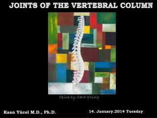
Joints of the Vertebral Column Atlanto-Occipital Joints The atlanto-occipital joints are synovial joints that are formed between the occipital condyles, above the facets on the superior surfaces of the lateral masses of the atlas. They are enclosed by a capsule. Ligaments Anterior atlanto-occipital membrane Posterior atlanto-occipital membrane Movements Flexion, extension, and lateral flexion. No rotation is possible
Atlantoaxial Joints The atlantoaxial joints are three synovial joints: one is between the odontoidprocessand the anterior arch of the atlas the other two are between the lateral masses of the bones The joints are enclosed by capsules. Ligaments Apical ligament: connects the apex of the odontoid process to the anterior margin of the foramen magnum. Alar ligaments: Cruciate ligament: This ligament consists of a transverse part and a vertical part. Membranatectoria: This is an upward continuation of the posterior longitudinal ligament. Movements There can be extensive rotation of the atlas and thus of the head on the axis.
Joints Between Two Vertebral Bodies The upper and lower surfaces of the bodies of adjacent vertebrae are covered by thin plates of hyaline cartilage. Sandwiched between the plates of hyaline cartilage is an intervertebral disc of fibrocartilage The collagen fibers of the disc strongly unite the bodies of the two vertebrae. Ligaments The anterior and posterior longitudinal ligaments run as continuous bands down the anterior and posterior surfaces of the vertebral column from the skull to the sacrum The anterior ligament is wide and is strongly attached to the front and sides of the vertebral bodies and to the intervertebral discs. The posterior ligament is weak and narrow and is attached to the posterior borders of the discs. These ligaments hold the vertebrae firmly together but at the same time permit asmall amount of movement to take place between them.
Joints Between Two Vertebral Arches The joints between two vertebral arches consist of synovial joints between the superior and inferior articular processes of adjacent vertebrae The articular facets are covered with hyaline cartilage, and the joints are surrounded by a capsular ligament. Ligaments 1-Supraspinous ligament: This runs between the tips of adjacent spines. 2-Interspinous ligament: This connects adjacent spines. 3-Intertransverse ligaments: These run between adjacent transverse processes. 5-Ligamentum flavum: This connects the laminae of adjacent vertebrae. In the cervical region, the supraspinous and interspinous ligaments are greatly thickened to form the strong ligamentum nuchae. The latter extends from the spine of the seventh cervical vertebra to the external occipital protuberance of the skull
Nerve Supply of Vertebral Joints The joints between the vertebral bodies are innervated by the small meningeal branches of each spinal nerve The nerve arises from the spinal nerve as it exits from the intervertebral foramen. It then re-enters the vertebral canal through the intervertebral foramen and supplies the meninges, ligaments, and intervertebral discs. The joints between the articular processes are innervated by branches from the posterior rami of the spinal nerves Read only
Muscles of the Back The muscles of the back may be divided into three groups: 1-The superficial muscles: connected with the shoulder girdle. 2-The intermediate muscles: involved with movements of the thoracic cage. 3-The deep muscles or postvertebral muscles belonging to the vertebral column • Deep Muscles of the Back (Postvertebral Muscles) • In the standing position, • the line of gravity passes through the odontoid process of the axis, • behind the centers of the hip joints, • and in front of the knee and ankle joints • thus, greater part of the body weight falls • in front of the vertebral column. • It is, therefore, not surprising to find that the postvertebral muscles of the back are well developed in humans. • The postural tone of these muscles is the major factor responsible for the maintenance of the normal curves of the vertebral column.
Superficial Vertically Running Muscles Erector spinae muscle Iliocostalis Longissimus spinalis • Intermediate Oblique Running Muscles • Transversospinalis: • SEMISPINALIS • MULTIFUDUS • ROTATORS
Vertebral column movement The following movements are possible: flexion, extension, lateral flexion, rotation, and circumduction. Flexion is a forward movement Extension is a backwardmovement Both are extensive in the cervical and lumbar regions but restricted in the thoracic region. Lateral flexion is the bending of the body to one or the other side. It is extensive in the cervical and lumbar regions but restricted in the thoracic region. Rotationis a twisting of the vertebral column. This is least extensive in the lumbar region. Circumduction is a combination of all these movements.