Download
1 / 45
810 likes | 2.52k Views
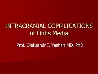
INTRACRANIAL COMPLICATIONS of Otitis Media. Prof. Oleksandr I. Yashan MD, PhD. Anatomy of the ear. Anatomy. Eust tube Petous apex. ME. Site of Ext Canal Mastoid. Carotid A. IAM. Cochlear aquiduct Endolymphatic sac & duct. Lateral sinus. M.E. Ext. A. M. Petrositis. Facial palsy.

E N D
INTRACRANIAL COMPLICATIONS of Otitis Media Prof. Oleksandr I. Yashan MD, PhD
Anatomy Eust tube Petous apex ME Site of Ext Canal Mastoid Carotid A. IAM Cochlear aquiduct Endolymphatic sac & duct Lateral sinus
M.E. Ext. A. M. Petrositis Facial palsy Mastoiditis Labyrinthitis Cranial Complications
M.E. Trigiminal gang Abducent VI th N Ext. A. M. Mastoid Pertrositis GradinigoSyndrome: Face pain (trigiminal neuralgia) Diplopia (VIth N) Increasing ear discharge
Abducent (VI th N) Palsy Abducent paralysis Normal
Facial Paralysis Inability to raise eye brow (frontalis m.) Inability to close eye lids (orbicularis occuli) Deviation of the mouth angle to opposite side (orbicularis oris) Failure to blow the mouth or build intra-oral pressure on the affected side (buccinator m.)
OTOGENIC INTRACRANIAL COMPLICATIONS The extension of an infection from the ear to intracranial spaces of a skull. In such cases the severe, sometimes fatal, diseases can develop.
OTOGENIC INTRACRANIAL COMPLICATIONS • Meningitis; • Abscess of a brain or cerebella; • Sigmoid sinus thrombosis (and otogenic sepsis); • Subdural abscess; • Extradural abscess; • Arachnoiditis of posterior cranial fossa
THE REASONS of OTOGENIC INTRACRANIAL COMPLICATIONS • Acute suporative (purulent) otitis media; • Chronic otitis media; • Mastoiditis; • Labyrinthitis.
AGENTS of OTOGENIC INTRACRANIAL COMPLICATIONS • Viruses; • Staphylococcus; • Streptococcus; • Pneumococcus - and dyplococcus; • Protey; • Pseudomonas aerogenoza; • Funguses; • Combination of microorganisms.
Paths of penetration of an infection to the intracranial spaces • Contact (or on a prolongation), through the holes in the bony walls of middle and inner ear (due to destructions) or through dehiscence in walls of temporal bone arises immediate contact inflammatory of the center with brain shells. • Haemotogenic or lymphogenic, when the infection penetrates intracranial space from a blood or lymph. • Through the prephormed paths – peryneural or peri vascular when inflammatory the process is spreaded to intracranial space through channels, that naturally exist in temporal bone (where nerves, vessel pass) and dehiscence in walls of middle ear. • Labyrinthogenic - through aqueducts of the inner ear.
Pathogenesis of OICC • Impairment of exudates drainage from middle or inner ear; • Eustachian tube not capable to take out the exudates from the ear to nasopharynx, and outflow it through a tympanic membrane is hampered. • Exudates or cholesteatoma are accumulated in a tympanic cavity or mastoid process’s cells, where the pressure is increased. That promotes a penetration of infection by different ways into intracranial space.
Pathogenesis of OICC • Due to contact mode of a penetration the infection to the intracranial space, the accumulation of pus or cholesteatoma in attic or antrum results in destruction of the upper wall of a tympanic cavity or antrum. Contaminated exudate spreads under a dura mater of middle cranial fossa, where extradural abscess is created. • In a place of abscess dura mater varies, becomes penetrable for toxins and bacterias, which hitting in space between dura mater and arachnoidea, forms subdural abscess. The more deep penetration of infection in brain substance finishes in creation of brain abscess
Erosion of tegmen, extension of infection below dura Brain Dura Tegmen Attic Antrum Tymp. Memb. (TM) Lateral sinus Mastoid Extra-dural Abscess Cholesteatoma & Attic perf or Central Perf
Erosion of tegmen, extension of infection below dura= Extradural Abscess Erosion of the dura and extension to lepto-meningies (Pia & Arachenoid mater = meningitis Meningitis
Perisinus abscess Brain Dura Tegmen Attic Cholesteatoma Lateral sinus Sinus plate Extension of infection inside the sinus cause first partially occluding infected thrombus Erosion of sinus plate and extension of infection above sinus dura = Peri-sinus Abscess The thrombus occludes the sinus and extends up & down Lateral Sinus Thrombosis
Lat. Sinus thrombosis Mast. Emissary V. & Griesinger sign Super. & inf. Petrosal sinuses Cavernous S. Int. Jugular. V. Extension of thrombus
Temporal Lobe Cereb- ellum Brain Abscess Direct extension of infection through meningies Indirect vascular extension Temporal Lobe Cereb- ellum
Temporal Lobe Abscess المناطق التوافقية (مناطق البيان والفقه) منطقة فقه القراءة (الفقه البصرى) منطقة البيان الكتابى Motor area Sensory area منطقة بروكاس: البيان الكلامى منطقة فقه الكلام (الفقه السمعى)
Pathogenesis of OICC • If inflammatory process penetrates in the skull: either through a posterior-internal wall of mastoid process, or through internal auditory canal, or through the aqueducts of inner ear. The inflammation process progresses in posterior cranial fossa with formation of perysinuses abscess or cerebella abscess. • The generalization of process, due to reduced resistance of macroorganism and high resistance of micro flora, develops meningitis, meningoencephalitis or sepsis. All intracranial complications can be mortal.
The doctors, which have taken part in diagnosis and in treatment of the patient with ICC: • ENT- physician; • Reanimation physician; • Neuropathology physician; • Neurosurgery physician; • Therapy - physician; • Ophthalmology physician; • Infection physician; • Phtysiatry - physician; • X-ray physician.
OTOGENIC MENINGITIS • Inflammation of all brain shells otologyc origin . • Can be serous or purulent
OTOGENIC MENINGITIS Meningeal symptoms • Appears due to tension of spinal nerve roots to the dura mater. They are: • Rigidity of neck muscles - impossibility to get by a patient’s chin to physician’s thumb located on jugular fossa; • Kerning - impossibility to unbend a leg in a knee joint after simultaneous bending of knee and coccyx joints; • Brudzinsky upper – when patient try to get by the chin to jugular fossa the patient’s knees are reduced to a stomach; • Brudzinsky middle – during pressing by physician’s fists on patient’s symphis the knees are reduced to the stomach; • Brudzinsky lower - during checking Kerning symptom - other leg are reduced to the stomach.
OTOGENIC MENINGITIS ІІ. Brain symptoms • Appears due to raising of spinal liquor pressure: • Deterioration of consciousness (sopor, stupor or coma). • Severe headache, • Nausea, vomiting, • hyper aesthesia (light phobia, noise phobia, raising of tactile and temperature sensitivity). • Forced position of the patient: „ Raising cock” pose;
OTOGENIC MENINGITIS The common – intoxication symptoms • Fever, temperature curve - constant; • Tachycardia; • Tachypnoe; • Blood hypertension • Hypo or anuria.
Additional methods of inspection • Blood analysis: neutrophillic leycocytosis, accelerated SОЕ is expressed; • Spinal punction - liquid implies under the increased pressure, it is muddy, and also contains to much cells elements, protein, sometimes - bacteria, contents of sugar and chlorides are reduced. • Bacteriological research of ear pus and spinal canal liquid shows the agent and its antibiotics resistance (to choice the adequate antibiotic therapy). • X-ray of temporal bone. • Computer tomography and magnet -nuclear resonance research - detection of cavities, filled with exudates in temporal bone.
Treatment • Urgent hospitalization in ЕNT -department, where urgent surgical procedure will performed (extended radical mastoidectomy). From usual mastoidectomy it differs by maximum wide disclosure of a dura mater of middle and posterior cranial fosses, and available mastoid cells. Postauricular wound is not closed to give possibility of cleaning of operational cavity and introduction medicines in operational wound. • The intensive conservative therapy includes: treatment by antibiotics in maximum dozes, that penetrate through blood-brain barrier (including in spinal canal), massive des intoxication, de hydratation, hormones.
ABSCESS of temporal lobe or CEREBELLA • Suppurative inflammation of brain substance, which localized mainly in cerebellum or in temporal lobe. • Can be multiple abscesses. • Quite often caused by anaerobic infection
STAGES of ABSCESS of the BRAIN and CEREBELLA • 1. The initial stage (2 weeks) is characterized by insignificant changing of the general patient’s, subfebrile temperature, increasing of headache on pathologic side, possible nausea and vomiting. The clinical patient’s state is like accution of chronic suppurative otitis media. • 2. In latent stage (2 weeks) the general state is changes unsignificantly. Depression, occasionally - diffuse or local headache. Tiredness, sleepiness, lack of appetite. Temperature may be normal.
STAGES of ABSCESS of the BRAIN and CEREBELLA • 3. The transformation in manifest stage happens gradually or suddenly. Presentation of arising of mental changing: depression, apathy, dormancy, slow silent language, attack of sleepiness at preservation of consciousness. Also: diminution of a patient’s mass, vomiting, local painfulness during percussion on skull, lack of appetite, smell from a mouth. Temperature can be normal or subfebrile, deceleration of a rhythm of intimate reductions (bradycardia), leukocytosis. Neurological manifestation of the brain abscess is a paralysis of arm or leg on opposite side. In this stage in majorities of cases it is possible to do the previous diagnosis. • 4. The terminal stage is characterized by coma, which develops due to progressing encephalitis, brain edema, diffuse meningitis or breaking of abscess in the cerebrum ventricles.
LOCAL sings of temporal lobe abscess • Amnesty aphasia (Right hand patients with abscess, located in the left brain lobe, can not term noun), • Motor aphasia (the patients can understand the language, but can not speak) • Sensory aphasia (the patients lose a possibility to understand the people’s language and himself language). • Abscess in the not conducting brain lobe is exhibited by mental disorders – lose of criticism, euphoria or depression. That can be imperceptible.
LOCAL sings of CEREBELLA ABSCESS • General patient’s state is more severe, than in patient with temporal lobe abscess. Rough rotatoric nistagmus directed to a pathologic side. • Significant deterioration of an equilibrium and coordination. • For diagnostics of cerebella abscess is necessary to fulfill a finger – nose, finger-finger, or tests, the patient misses out only by the hand on a side of abscess disposition. • In tests on an equilibrium: Romberg pose - direction of a falling does not vary at position of the head; at a straight gate patient with the closed eyes the course deviates in the abscess side. Besides the sign cerebella abscess is positive test on adiadochokinetic (hand on pathologic side defeats) and muscles hypotonia on abscess side.
Additional inspection method • Common blood analysis: possible leukocyteosis, accelerated SОЕ; • Spinal punction - liquid implies under the raised pressure, transparent, increased contents of proteins and insignificant pleocytosis. • Computer tomography imaging and magnet -nuclear resonance research - allows to reveal the abscess in the substance of brain, and to estimate it size and localization. • Bacteriological research of ear pus and spinal canal liquid shows the agent and its antibiotics resistance (to choice the adequate antibiotic therapy). • Echoencephalographia - dislocation of brain structures
Treatment • Surgical disclosure of the purulent center in temporal bone together with wide disclosure of a dura mater of middle or posterior cranial fossa (extended mastiodectomy or extended radical operation on the ear). • Through an operational wound make a injection of brain substance on depth in 4 cm with the purpose of searching abscess. Its uncovering and drainage to maintain constant outflow and washing of the abscess cavity until it full obliteration. • After operation the patient translate in reanimation department, where continue intensive conservative therapy: treatment by antibiotics, that penetrate through hematological-encephalitic barrier, in maximum dozes (including in spinal canal), massive des intoxication, de hydratation, hormones.
SINUS THROMBOSIS And OTOGENIC SEPSIS • Sinus thrombosis arises mainly due to at mastoiditis and epitympanitis. The accumulation of pus in air cells located near sigmoid sinus, reduces in destruction inner bony of a wall mastois and creation pery sinus abscess. • In a place abscess the sigmoid sinus wall inflammated - develops peryphlebitis. Afterwards inflammatory the process passes on inner surface of sinus (endophlebitis). In a sinus lumen wall thromb is created, it gradually increases in sizes and obstructs the sinus. Due to inflammation thromb purulently fuses, and it contaminated parts by the blood current are delivered on all organism (septicopiemia or septicehaemy), creating the lesions of remote metastases (in the lungs, muscles, joints, interior organs and so on).
CLINIC of SINUS THROMBOSIS • Sever common condition of the patient, is exhibited first of all by deterioration of central nervous system functions (stupor, coma), septic body temperature (more then 1 day differential), ague, dense sweat, tachycardia, painfulness of mastoid process, magnification and painfulness of neck lymphatic nods, accelerated SОЕ. • Sometimes on the foreground appear septic lesions of interior organs: lungs, heart, kidneys, liver, digestive tract and so on. Hemorrhages in interior organs sometime happen, also under the skin and mucous lining. The skin quite often gains yellow coloring
Additional inspection methods • Common blood analysis: possible leukocyteosis, accelerated SОЕ; • Spinal punction - liquid implies under the raised pressure, transparent, increased contents of proteins and insignificant pleocytosis. • Computer tomography imaging and magnet -nuclear resonance research - allows to reveal the inflammatory lesion near or in the sigmoid sinus, and presence of thromb in it lumen. • Bacteriological research of ear pus and spinal canal liquid shows the agent and its antibiotics resistance (to choice the adequate antibiotic therapy). • Echoencephalographia- dislocation of brain structures.
Treatment • In suspicion of sinus thrombosis the patient must be urgently transported to ЕNТ-department. After installation of the diagnosis it is necessary to perform immediately operation. In patient with acute otitis media the extended mastoidectomy is necessary, in patient with chronic suppurative otitis media - extended radical mastoidectomy operation on ear. The aim of surgical interference is to remove inflammatory lesions in temporal bone, to disclose the sigmoid sinus, and to remove obliterative thromb (if persist). • Intensive conservative therapy: treatment by antibiotics, that penetrate through hematological-encephalitic barrier, in maximum dozes (including in spinal canal), massive des intoxication, de hydratation, hormones and anticoagulants. • Prognosis favorable at opportune and combined (surgical and conservative) treatment.
After-care on the patients with intracranial complications • After-care on the patients with intracranial complications has especially important value. The general state of the patient is commonly very severe and requires singular attention of medical staff. A right after-care has not smaller value, than surgical treatment, in preservation of patient’s life. • Such the patient is necessary to put in separate small chambers with the blacked out windows (light phobia), to ensure full silence (noise phobia), comfortable temperature and strict bed condition. • Is necessary to ensure constant supervision of the general patient’s state, his consciousness, temperature, palpitation, and these data in an hour are spelled in the temperature leaf. Transport such patients (on bandagings) only on wheeled bed.
Prophylaxis of intracranial complications • Consists in opportune detection both right treatment of acute and chronic suppurative otitis media. • Mandatory operating treatment in cholesteatoma; • Early paracentesis of a tympanic membrane in patient with acute purulent otitis media