Cell Division
Cell Division. Biology Mr. Greene Unit 7. Bellringer. Each time a cell reproduces, it divides into two new cells. When each of the new cells divide, the result is four new cells. If this continues, how many cells will be present after the cells reproduce 6 times?. Key Ideas.


Cell Division
E N D
Presentation Transcript
Cell Division Biology Mr. Greene Unit 7
Bellringer Each time a cell reproduces, it divides into two new cells. When each of the new cells divide, the result is four new cells. If this continues, how many cells will be present after the cells reproduce 6 times?
Key Ideas • Why do cells divide? • How is DNA packaged into the nucleus? • How do cells prepare for division?
webbed fingers Why Cells Reproduce • As the body of a multicellular organism grows larger, its cells do not also grow large. Instead, the body grows by producing more cells. • New cells are needed to help tissues and organs grow. • As old cells die and new cells take their place. • New cells also replace damaged cells.
Why Cells Reproduce, continued Cell Size • A cell grows larger by building more cell products. To do this, the cell must take in more nutrients, process them, and get rid of wastes. • A cell’s ability to exchange substances is limited by its surface area–to-volume ratio. As a cell gets larger, substances must travel farther to reach where they are needed.
Why Cells Reproduce, continued Cell Maintenance • The work of cells is done by proteins. As a cell gets larger, more proteins are required to maintain its function. • If the cell gets too large, DNA instructions cannot be copied quickly enough to make the proteins that the cell needs to support itself. • Cell size is also limited by the cell’s DNA.
How Small Is a Cell? • it varies • the egg cell is 14 times as large as a red blood cell • the contents of cells cannot be too far from the membrane. • i.e. responses would take too long if cells were too big • the smaller the cell, the more efficient it becomes • i.e. look at the surface area to volume ratio
Ratio of Surface Area to Volume in Cells Cell Size Surface Area (l x w x 6) Volume (l x w x h) Ratio of Surface Area to Volume
Why Cells Reproduce, continued Making New Cells • Each “daughter” cell has a higher surface area–to-volume ratio than its parent does. • Each new cell also gets an entire copy of the cell’s DNA. • Because larger cells are more difficult to maintain, cells divide when they grow to a certain size.
Chromosomes • The large molecule of DNA is organized into hereditary units called genes. • A gene is a segment of DNA that codes for RNA and protein. • Each cell has a large amount of DNA that must be condensed into a very small volume. • DNA is organized and packaged into structures called chromosomes.
Chromosome Structure • it is a tightly coiled mass around proteins • not visible because it is spread throughout the nucleus • chromosomes - tightly compacted bodies of DNA which arise before cell division (mitosis) • chromatids - copies of each chromosome which form just prior to division • centromere - protein disk which hold the chromatids together
a set of chromosomes is called a karyotype there are 46 chromosomes in a single human cell you can sort them out into 23 pairs by size and shape homologous pairs = similar in shape and size and information. The pairs are almost identical copies of one another diploid(2n) - two copies of chromosomes haploid(n) - one copy of chromosomes Gamete cells (sperm and egg) have 1 homologue. Chromosome # in Organisms mosquito 6 fly 12 frog 26 mouse 40 human 46 chimpanzee 48 orangutan 48 gorilla 48 horse 64 dog 78 duck 80 Chromosome Number
Sex Chromosomes • sex chromosomes • the pair which determines if you are a male or female • Female = XX • Male = XY • If you are male, you received your Y from your father. • The sex chromosomes are separated during meiosis.
parent cell DNA duplicates cell begins to divide daughter cells Chromosomes, continued • A prokaryotic cell has a single circular molecule of DNA. • This loop of DNA contains thousands of genes. • A prokaryotic chromosome is condensed through repeated twisting or winding, like a rubber band twisted upon itself many times.
chromatid telomere centromere telomere Condensed, duplicated chromosome Chromosomes, continued Eukaryotic Chromosomes • Eukaryotic cells contain many more genes arranged on several linear DNA molecules. • Eukaryotic DNA into highly condensed chromosome structures with the help of many proteins. • The DNA and proteins make up a substance called chromatin.
SupercoiledDNA DNA andhistones DNA doublehelix Chromatin Chromosomes, continued Eukaryotic Chromosomes • The first level of packaging is done by a class of proteins called histones. A group of eight histones come together to form a disc-shaped histone core. • The long DNA molecule is wound around a series of histone cores in a regular manner and is called a nucleosome. Under an electron microscope, this level of packaging resembles beads on a string. • The string of nucleosomes line up in a spiral to form a cord that is 30 nm in diameter.
Chromosomes, continued Eukaryotic Chromosomes • During most of a cell’s life, its chromosomes exist as coiled or uncoiled nucleosomes. • As the cell prepares to divide, the chromosomes condense even further ensuring that the extremely long DNA molecules do not get tangled up during cell division.
Chromosomes, continued Eukaryotic Chromosomes • The nucleosome cord forms loops that are attached to a protein scaffold. These looped domains then coil into the final, most highly condensed form of the chromosome. • Many dense loops of chromatin form the rod-shaped structures that can be seen in regular light microscopes. • Each of the two thick strands of a fully condensed, duplicated chromosome are called a chromatid. • Each chromatid is made of a single, long molecule of DNA.
Chromosomes, continued Eukaryotic Chromosomes • Identical pairs, called sister chromatids, are held together at a region called the centromere. • During cell division, the sister chromatids are separated at the centromere, and one ends up in each daughter cell. • Each new cell has the same genetic information as the parent cell.
Preparing for Cell Division • All new cells are produced by the division of preexisting cells. • The process of cell division involves more than cutting a cell into two pieces. Each new cell must have all of the equipment needed to stay alive. • All newly-formed cells require DNA, so before a cell divides, a copy of DNA is made for each daughter cell. • Each new cells will function in the same way as the cells that they replace.
Preparing for Cell Division, continued Prokaryotes • In prokaryotic cells, the circular DNA molecule is attached to the inner cell membrane. • The cytoplasm is divided when a new cell membrane forms between the two DNA copies. Meanwhile the cell continues to grow until it nearly doubles in size. Prokaryotes • The cell is constricted in the middle, like a long balloon being squeezed near the center. • Eventually the dividing prokaryote is pinched into two independent daughter cells, each of which has its own circular DNA molecule.
Preparing for Cell Division, continued Eukaryotes • The reproduction eukaryotic cells is more complex than that of prokaryotic cells. • Eukaryotic cells have many organelles. In order to form two living cells, each daughter cell must contain enough of each organelle to carry out its functions. • The DNA within the nucleus must also be copied, sorted, and separated.
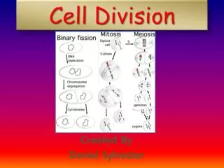
Visual Concept: Comparing Cell Division in Prokaryotes and Eukaryotes Click above to play the video.
Summary • Because larger cells are more difficult to maintain, cells divide when they grow to a certain size. • Many proteins help package eukaryotic DNA into highly condensed chromosome structures. • All newly-formed cells require DNA, so before a cell divides, a copy of its DNA is made for each daughter cell.
Bellringer Using the diagram of a football field, write a short paragraph describing how the players, the midfield line, and the goal posts compare to the structures of a cell involved in mitosis and cell division.
Key Ideas • What are the phases of the eukaryotic cell cycle? • What are the four stages of mitosis? • How does cytokinesis occur?
Cell Division • karyokinesis - the division of nuclear material • mitosis - the process by which the nucleus of a eukaryotic cell divides to form two nuclei
M phase (Mitosis) Interphase G1 phase S phase G2 phase Prophase Metaphase Anaphase Telophase Concept Map Cell Cycle includes is divided into is divided into
Eukaryotic Cell Cycle • The cell cycle is a repeating sequence of cellular growth and division during the life of a cell. • The cell cycle is made up of five phases. The first three phases together are known as interphase. The remaining two phases make up cell division.
Interphase Interphase – The longest part of a cells life Three subphases G1 - first gap cell undergoes intense growth longest phase for most cells synthesize proteins S phase - synthesis copies of DNA are made 4 copies of each chromosome G2 - second gap usually the shortest of the 3 phases preparations for cell division duplicates its organelles nerve cells are arrested in the G1 phase referred to as G0 fibroblasts (heal wounds) grow on demand, otherwise quiescent
Visual Concept: Cell Cycle—G1 Phase Click above to play the video.
Visual Concept: Cell Cycle—S Phase Click above to play the video.
Visual Concept: Cell Cycle—G2 Phase Click above to play the video.
Visual Concept: Cell Cycle—M Phase Click above to play the video.
Visual Concept: Mitosis Click above to play the video
Mitosis – Stage 1 - Prophase first and longest phase of mitosis chromosomes coil into short rods = chromatids (sister) nuclear envelope breaks up centrioles form two tiny structures located in the cytoplasm near the nuclear envelope that separate and take up positions on opposite sides of the nucleus. spindle fibers - protein cables - form across the cell and enter the nucleus
Prophase Click to animate the image. C B F E A D
Mitosis – Stage 2 - Metaphase often only lasts a few minutes chromosomes attach to the spindle fibers at the kinetochores chromosomes line up in the center of the cell equatorial plane
Mitosis – Stage 3 - Anaphase shortest phase driven by microtubules each chromosome separates from its identical copy chromosomes are reeled to opposite sides of the cell spindle fibers begin to break down
Mitosis – Stage 4 - Telophase each side now has a complete set of chromosomes nuclear envelope now develops around each new set of chromosomes chromosomes uncoil spindle fibers disappear
Finishing Up the Division CYTOKINESIS Mitosis is followed by cytokinesis - cell division the cell divides into two cells with its own nucleus Animal Cells the cell membrane is drawn inward until the cytoplasm is pinched in half (cleavage furrow) forming two new cells (daughter cells) Plant Cells plant cells develop a cell plate midway between the 2 new ones the cell wall then begins to appear in the cell plate sometimes karyokinesis occurs without cytokinesis and multinucleated cells result
The Cell Cycle G1 phase M phase S phase G2 phase
Visual Concept: Comparing Cell Division in Plants and Animals