
Development of a Biofeedback Training System for Enhanced Athletic Performance
This project focuses on creating a biofeedback training system to enhance athletic performance. By utilizing electromyography to measure electrical signals from muscle contractions, the system aims to provide real-time feedback on muscle fatigue. A microcontroller processes these signals, determining when muscle fatigue threshold is reached and offering responses to the user. This system can benefit individuals of all ages and fitness levels, assisting them in maximizing exercise benefits, reaching goals faster, and improving overall health. Continuous enhancements are being made for accurate muscle fatigue measurement.

Development of a Biofeedback Training System for Enhanced Athletic Performance
E N D
Presentation Transcript
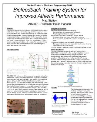
Abstract: The goal of this project is to produce a biofeedback training system that leads to improved athletic training. Electrical signals produced by muscles when they contract, measured by an electromyograph, are used as an indicator of muscle fatigue. The measured signals are processed using a microcontroller, which analyzes the signals and provides a feedback response to the user when the amplitude gets below a certain threshold. In the end, a biofeedback training system was produced that can help people of all ages and fitness levels maximize their benefits from exercise, reach their goals faster, and improve their health. • Design Requirements: • Measure electrical signals from muscles • Use electrodes to measure electrical signals • Amplify signal so it can be analyzed • Analyze signal to determine level of muscle fatigue • Analog-to-digital conversion of signal from muscle • Measure maximum and relative maximum amplitude peaks of signal • Determine threshold at which muscle fatigue occurs • Compare relative maximum to absolute maximum of signal to determine level of muscle fatigue • Provide feedback response to user when muscle fatigue occurs Senior Project – Electrical Engineering- 2009Biofeedback Training System for Improved Athletic PerformanceMatt StattonAdvisor – Professor Helen Hanson Signal Processing: The electromyogram was analyzed using a Silicon Labs C8051F020 microcontroller. After analog-to-digital conversion using the on-board 12-bit analog-to-digital converter, the microcontroller was programmed to measure the average amplitude of the signal during the first repetition. A running average of the following repetitions was compared to the initial average, and a feedback response was provided to the user when the running average reached a threshold of muscle fatigue based on the initial average. Electromyograph: Figure 1: EagleCAD schematic designed based on circuit diagram from http://instruct1.cit.cornell.edu/courses/ee476/FinalProjects/s2005/bsm24_ajg47/website/website/index.html Figure 2: EagleCAD board file created from schematic A MAX666CPA voltage regulator was used to regulate voltage from a 9V battery to 5V to power the operational amplifiers. An LT1494 operational amplifier, with gain A = 1, was used to create a virtual ground of VCC/2 = 2.5 V. An INA106 differential amplifier, with internal gain A = 10, was used to amplify the differential input from two surface electrodes placed on the proximal and distal portions of the muscle. External resistors were used to increase the gain to A = 110. The electromyograph was created using an LPKF ProtoMat C20S circuit board plotter from a schematic made in EagleCAD. All parts were soldered into the board created by the circuit board plotter. The inputs were connected to electrode leadwires, and the output was connected to the 8051 microcontroller for processing. Figure 5: Flow chart of electromyograph signal analysis program Results: The electromyograph measured the electrical activity of the muscles. However, the microcontroller has not yet been able to consistently measure muscle fatigue based on the amplified electromyogram data. Further work is being done to enable the microcontroller to more accurately measure the level of muscle fatigue of the user. Figure 6: Measured Electromyogram signal displaying fully contracted, minimally contracted, and resting biceps muscle Acknowledgments: Professor Hanson Professor Hedrick Ben Bunes Internal Education Fund Figure 3: Front of electromyograph circuit Figure 4: Back of electromyograph circuit


