Download
1 / 36
450 likes | 1.14k Views
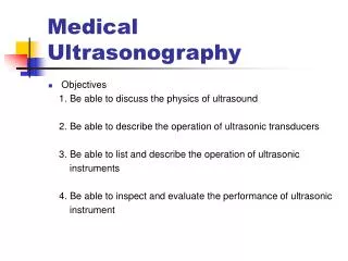
Medical Ultrasonography. Objectives 1. Be able to discuss the physics of ultrasound 2. Be able to describe the operation of ultrasonic transducers 3. Be able to list and describe the operation of ultrasonic instruments

E N D
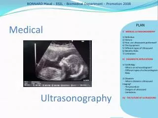
Medical Ultrasonography • Objectives 1. Be able to discuss the physics of ultrasound 2. Be able to describe the operation of ultrasonic transducers 3. Be able to list and describe the operation of ultrasonic instruments 4. Be able to inspect and evaluate the performance of ultrasonic instrument
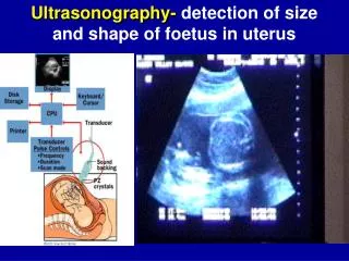
17-3 What is ultrasound ? • Medical ultrasound systems operate at frequencies of up to 10MHz or more • An ultrasonic wave is acoustical:is a mechanical wave in a gaseous,liquid,or solid medium • The mechanical wave consists of alternating areas of higher and lower pressure
Distinguish with the radio signals and the ultrasound signals • Radio signals: are electromagnetic waves i.e: If an alternating current(ac) oscillation of,say,2500kHz,were connect to an appropriate antenna,then radio would be launched. • Ultrasound signals: are acoustical i.e: If that same 2500kHz ac signal were applied to an ultrasound transducer,then an acoustical signal would be launched.
17-4 Physics of sound and ultrasound waves • In radio signals:thevelocity is about 300,000,000m/s • In ultrasound signals:the velocity =
17-4-2 Reflection,refraction,diffraction,and scattering phenomena • Reflection and refraction of waves
Diffraction:is a bending of the direction of propagation that occurs when a wave impinges on an object of different density embedded within,and surrounded by,the incident media. diffraction: can distort the direction information of the signal.
17-4-3 Specular reflection,diffuse reflection,and scattering
17-4-4 Acoustical impedance • Acoustical impedance:is a measurement of its opposition to the propagation of sound waves
17-5 Ultrasound transducer • In ultrasound,two transducer functions are recognized: (1) conversion of ac oscillations into acoustical vibratioon (2) conversion of acoustical oscillations back into electrical oscillations These two functions are the transmit and receive transducers
Inverse square law • Inverse square law:the power density drops according to the square of the distance
The resolution of the ultrasonic signal • The pulse width: sets the maximum longitudinal resolution also called range resolution • The beamwidth: sets sets the azimuthal or angular resolution
17-6 Absorption and attenuation of ultrasound energy • Look the eq(17-9) in the textbook • The value of attenuation by absorption depends on both the acoustical impedance and the frequency used. • Figure 17-12 shows how soft-tissue absorption losses rise with frequency
17-7 Scan modes and scanning systems • A-scan mode: uses a stationary transducer to fire a pulse into tissue
B-scan • B-scan mode: use the same time base as the A-scan but but plots the strength of the returning signal as changes in brightness i.e: a strong reflection is brighter than the weaker reflection
Time-motion(T-M) mode • M-mode: is essentially an A-scan but with successive looks at the target created by scanning the time base vertically. look at the figure17-19
17-7-1 Electronically scanned phased array transducers • The ESPA angle = eq 17-12 • The ESPA combined output voltage = eq 17-13
ESPA’s grating lobes problem • The angle at which grating lobes appear = eq 17-14 • The grating lobes will cause spurious returns to to be displayed, some suppression will reduce this situation(especially signal strength display)
17-9 Doppler effect • Doppler effect: is a change of frequency that occurs when the receiver and transmitter move relative to each other • The frequency shift = eq 17-16
17-10 Transcutaneous Doppler flow detectors • If the blood were motionless,the frequency shift would be zero ,so the return wave frequency will be identical to the incident wave frequency. • But moving blood produces a shift that is proportional to the blood velocity. • A frequency shift of shift of approximately 200Hz at 10MHz corresponds to a blood velocity of approximately 6 cm/s.
17-11 Flowmeters • The average flow velocity = eq 17-17
Doppler flowmeter • This transducer uses the frequency change of a wave scattered from particulate matter flow in the vessel • Look at the eq 17-19
Summary 1 Medical ultrasound tends to use frequency in the 2-10MHz 2 Ultrasound waves obey the reflection ,refraction,diffraction,and scatter rules of ordinary wave behavior. 3 Several factors affect biological interaction with ultrasound :frequency,irradiation time,beam intensity,and duty cycle. 4 Most ultrasound transducer are piezoelectric crystals.