Download

1 / 41
651 likes | 2.06k Views
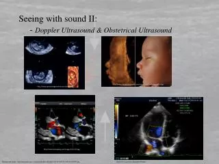
Doppler Ultrasonography in Obstetrical Practice. China Medical University Hospital OBS & GYN department Chien Chung, Lee.

E N D
Doppler Ultrasonography in Obstetrical Practice China Medical University Hospital OBS & GYN department Chien Chung, Lee
MaternalChronic hypertensionCollagen-vascular diseasesSickle cell anemiaCurrent substance abuseImpaired renal functionAsthmaPneumoniaSignificant cardiac diseaseSeizure disordersDiabetesAcute febrile illnessesSignificant anemia (hematocrit <26% FetalIntrauterine growth restrictionCongenital anomaliesFetal cardiac arrhythmiasIsoimmunizationHydrops fetalisFetal infections such as parvovirus, coxsackievirus B, syphilis, toxoplasmosis Conditions That Place Fetuses at Risk for Adverse Outcomes Pregnancy-relatedPoorly controlled gestational diabetesMultiple gestationsPregnancy-induced hypertensionCholestasis of pregnancyPremature rupture of the membranes (preterm)Unexplained elevated maternalserum alpha-fetoproteinPolyhydramniosOligohydramniosPlacental abruptionAbnormal placentationPostdatesUnexplained stillbirth in a prior pregnancy
Biophysical profile for fetal assessment in high risk pregnancies (Cochrane Methodology Review) Reviewers' conclusions: At present, there is not enough evidence from randomised trials to evaluate the use of biophysical profile as a test of fetal well-being in high risk pregnancies. Alfirevic Z, Neilson JP. In: The Cochrane Library, Issue 4, 2003.
Doppler ultrasound for fetal assessment in high risk pregnancies (Cochrane Methodology Review) Reviewers' conclusions: The use of Doppler ultrasound in high risk pregnancies appears to improve a number of obstetric care outcomes and appears promising in helping to reducing perinatal deaths. Neilson JP, Alfirevic Z.. In: The Cochrane Library, Issue 4, 2003.
Descriptive characteristics of randomized trials evaluating the use of Doppler ultrasonography in pregnancy included in overview
Proportional effect of Doppler ultrasonography on number of dead babies (stillbirths and neonates) when used in high-risk pregnancies. Meta-analysis shows that clinical action guided by Doppler ultrasonography reduced the odds ratio of perinatal death by 38%
Effects of Doppler ultrasonography on perinatal outcomes in high-risk pregnancies. Post hoc analysis. The 16% reduction in the number of elective deliveries, 31% reduction in fetal distress in labor, and 87% reduction in hypoxic encephalopathy in the Doppler group reached statistical significance.
Conditions for Doppler ultrasound • Pregnancies complicated by IUGR • Pregnancies in which the fetus is at risk for anemia • Multiple gestations • Pregnancies treated with prostaglandin inhibitors to monitor the ductus arteriosus • Fetal echocardiograms
Doppler Flow Velocity in the First Trimester Comparison of endometrial thickness, RI, & gestational age between groups Alcazar JL, Ortiz CA. Eur J Obstet Gynecol Reprod Biol. 2002 Apr 10;102(1):83-7.
Doppler Flow Velocity in Uterine Artery Bewley et. al. Br J Obstet Gynaecol 1989;96:1040–6
Normal uterine artery at 12 weeks shows relatively high resistance, absent notching. • Normal midtrimester uterine artery, increased diastolic flow. • Normal third trimester uterine artery, very low resistance. • High resistance with persistent notching may be normal in first trimester, not in this 24-week gestation. • Very high resistance, marked notching, absent diastolic velocities in a woman with pre-eclampsia, and severe intrauterine growth restriction (IUGR) at 28 weeks.
Doppler Flow Velocity in Umbilical Artery • Normal umbilical artery at 18 weeks shows relatively high resistance, but consistent diastolic flow. • Normal umbilical artery at 36 weeks, low resistance, generous diastolic flow. • High resistance, diastolic velocity low. • Absent end-diastolic velocity (AEDV). • Reversed diastolic velocity (REDV) in severe intrauterine growth restriction (IUGR).
Doppler Flow Velocity in Umbilical Artery Fetuses with absent end-diastolic velocity of the umbilical artery all died in utero within 3 weeks (median 7 days). Madazli R, Uludag S, Ocak V. Acta Obstet Gynecol Scand 2001; 80:702
FACTORS AFFECTING UMBILICAL ARTERY DOPPLER FLOW VELOCITY WAVEFORMS
Diagnostic efficacy of umbilical arterial Doppler in IUGR Author DI Prevalence Sensitivity Specificity PPV Fleischer S/D>3.0 16.8 78 83 49 Aruidini PI>1SD 30.7 60.8 73 50 Berkowitz S/D>3.0 25 55 92 73 Divon S/D>3.0 35.4 49 94 81 Gaziano S/D>4.0 9.4 79 66 79 Ott S/D>3.0 10.4 59 84 29 Maulik S/D>2.9 12.3 75 71 27 Lowery S/D>4.0 22.6 65 66 24 Lee S/D>3.0 15 91.7 68.7 84.6
Middle cerebral artery Doppler waveforms Normal flow of the Middle Cerebral Artery in 1º trimester Normal flow of the Middle Cerebral Artery in 2º and 3º trimester
Middle cerebral artery Doppler waveforms (A) Normal middle cerebral artery (MCA) at term - normal peak systolic velocity (58 cm/s), high resistance, low end-diastolic velocity. (B) ‘Brain sparing’ MCA - lower peak, much higher diastolic velocity suggests cerebrovasodilation. (C) Anemic fetus with retained high resistance, elevated peak systolic velocity (77 cm/s).
The upper panel represents the venous waveform, correlated with the EKG in the lower panel. A = atrial systole, S = ventricular systole, D = early ventricular diastole. The colored portions of the waveform represent the Tamx for atrial systole (gold), ventricular systole (red), and early ventricular diastole (blue). The yellow arrows represent the measurement of the peak velocity for ventricular systole and early ventricular diastole. The black arrow represents the peak velocity for atrial systole.
(A) Ductus venosus (DV) Doppler waveforms at 12 weeks gestation. (B) At 12 weeks gestation, an abnormal a-wave (a), correctly predicted anomalous pulmonary and systemic venous return, proven by fetal echocardiography at 24 weeks. (C) DV at 26 weeks, with 4-phase waveform. (1) atrial contraction (2) ventricular systole, (3) return (ascent) of the annulus (called the y-descent of the DV waveform), & (4) diastole. (D) Normal waveform from the middle hepatic vein which, is only a few millimeters from the DV.
Progressive changes in Doppler parameters in IUGR fetuses delivered for an abnormal Biophysical Profile Score.
Hemodynamic changes occurring in fetal arterial vessels during hypoxemia and acidemia induced by uteroplacental insufficiency
Fetal Systemic Vascular Responses in IUGRA/REDV, absent or reversed end-diastolic velocitiesHARMAN: Clin Obstet Gynecol, 46(4).December 2003.931-946
Aortic isthmus blood velocity waveform a) normal blood flow pattern in an uncomplicated pregnancy b) antegrade net blood flow (antegrade/retrograde ratio of 2.0) c) retrograde net blood flow with a corresponding value of 0.54 in pregnancies complicated by placental insufficiency. In the sagittal view of the fetus, the aortic arch and the location of the aortic isthmus (white triangle) are shown.
Coronary artery blood velocity waveform of a growth-restricted 32 week fetus (heart sparing effect).
Alfred Abuhamad et al. Contemporary Ob/Gyn May 1, 2003;48:56-73
Evaluation of fetal intrapartum hypoxia by middle cerebral & umbilical artery Doppler velocimetry with simultaneous cardiotocography & pulse oximetry During active labor the fetus maintains oxygen supply to the brain by redistributing blood flow. In cases of hypoxia this is feasible for only 2 min. Siristatidis C, Salamalekis E, Kassanos D, Loghis C, Creatsas GArch Gynecol Obstet. 2003 Nov 5
Spectral Doppler waveform of an A-A anastomosis with characteristic bidirectional, pulsatile flow.
Systematic Doppler EvaluationHARMAN: Clin Obstet Gynecol, Volume 46(4).December 2003.931
Which Doppler Tests Should be Performed? • Uterine arteries depict maternal vascular effects of the invading placenta • Umbilical artery Doppler reflects downstream placental vascular resistance • Middle cerebral artery changes begin when the redistribution of cardiac output reflects rising placental resistance • precordial veins illustrate fetal cardiac function
DIFFERENTIAL DIAGNOSIS OF OLIGOHYDRAMNIOS PPROM --- normal renal vessels, normal umbilical flow & normal filling of the bladder. Bilateral renal agenesis or dysplasia --- umbilical artery Doppler is normal, but no renal vessels & no bladder filling Severe hypoxia with IUGR --- fetal measurements are small for gestation, fetal heart looks dilated & the bowel is echogenic. Doppler demonstrates the presence of two renal arteries and absent or reversed end-diastolic frequencies in the umbilical arteries
Deficient placentation defined by notched uterine arteries Increased umbilical artery resistance with progression to AEDV/REDV Declining CPR, brain-sparing MCA As the arteriovenous ratio decline, ductus venosus abnormality begins Abnormal biophysical variables emerge Oligohydramnios and abnormal (non-reactive) fetal heart rate tracing Loss of fetal breathing movements, body movements and fetal tone
THE USE OF FETAL DOPPLER IN OBSTETRICSSociety of Obstetricians and Gynaecologists of Canada. No. 130, July 2003 1.Umbilical artery Doppler should be available for assessment of the fetal-placental circulation in pregnant women with suspected severe placental insufficiency. (I-A) 2. Depending on other clinical factors, reduced, absent, or reversed umbilical artery end-diastolic flow is an indication for enhanced fetal surveillance or delivery. If delivery is delayed to enhance fetal lung maturity with maternal administration of glucocorticoid, intensive fetal surveillance until delivery is suggested for those fetuses with reversed end-diastolic flow. (II-1B) 3. Umbilical artery Doppler should not be used as a screening tool in healthy pregnancies, as it has not been shown to be of value in this group. (I-A) 4. Umbilical venous double pulsations, in the presence of abnormal umbilical artery Doppler waveforms, necessitate a detailed assessment of fetal health status. (II-3B) 5. Measurement of the fetal middle cerebral arteryDoppler peak systolic flow velocity is a predictor of moderate or severe fetal anemia and can be used to avoid unnecessary invasive procedures in pregnancies complicated with red blood cell isoimmunization. (II-1A) 6. Since inaccurate information concerning fetal Doppler studies could lead to inappropriate clinical decisions, it is imperative that measurements be undertaken and interpreted by expert operators who are knowledgeable about the significance of Doppler changes and who practise appropriate techniques. (II-1A)
Conclusion • No single diagnostic modality can provide information complete enough to adequately address the complex nature of IUGR and its interacting fetal compensations and compromises • Management decisions based on Doppler data are gestational age dependent