Download
1 / 6
60 likes | 322 Views
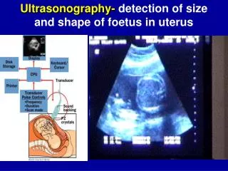
Ultrasonography- detection of size and shape of foetus in uterus. X-Ray Imaging methods : Sciascopy,Sciagraphy, Classical and Computer Tomography . X-rays: kind of ionizing, non-visible radiation.It is danger for living body . Max. harmless exposure is 5 mSv/ year

E N D
Ultrasonography-detection of size and shape of foetus in uterus
X-Ray Imaging methods:Sciascopy,Sciagraphy, Classical and Computer Tomography X-rays: kind of ionizing, non-visible radiation.It is danger for living body. Max. harmless exposureis5 mSv/ year Definition: is elmg. waving of photoelectrons (as a visible ligh), but with very short λ = 0.05 ηm Source: X-ray tube (diode with -Cathode and + Ano-de).Electrons are emitted from Cathode and flight in vacuum targeting Anode. Thus, only 1-2% X- ra-ys is produced, 98% is taken away of diode as a heat. The higher is anodal current (50-150 kV) the hardest (more penetrable) is X-ray radiation and vice ver-sa.X-ray photo is black-white.Thebones and air have best contrast, soft tissues have worse or no contrast.