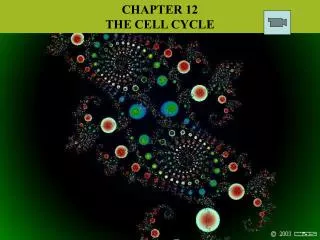
Chapter 12: The Cell Cycle
Chapter 12: The Cell Cycle. Cell division functions in reproduction, growth, and repair. Cell Division - the reproduction of cells Cell Cycle - the life of a cell from its origin in the division of a parent cell until its own division into two


Chapter 12: The Cell Cycle
E N D
Presentation Transcript
Cell division functions in reproduction, growth, and repair. Cell Division - the reproduction of cells Cell Cycle - the life of a cell from its origin in the division of a parent cell until its own division into two Cell division enables unicellular organisms to divide themselves, forming two separate organisms, and also enables sexually reproducing organisms to develop from a single cell- the fertilized egg, or zygote. After an organism is fully grown, cell division continues to function in renewal and repair.
Cell division distributes identical sets of chromosomes to daughter cells • A cell’s endowment of DNA is called its genome. Before a cell can divide, all of this DNA must be copied and then separated so that each daughter cell ends up with a complete genome. • During the replication and distribution of DNA, the DNA molecules are packed into chromosomes. Each eukaryotic species has a characteristic number of chromosomes in each nucleus. • Somatic cells – all body cells except the reproductive cells • Gametes – sperm and egg cells
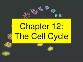
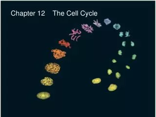
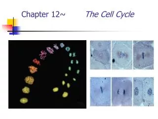
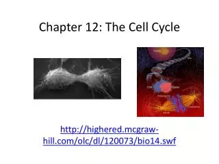
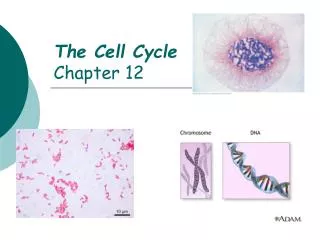
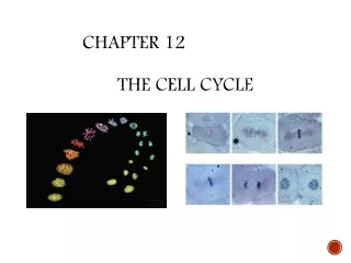
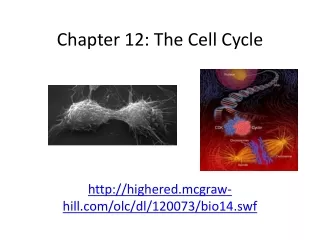
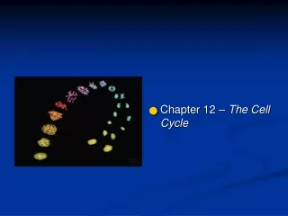
Chromosomes are duplicated and distributed in Mitosis, which isthe division of the cell’s nucleus • In each eukaryotic chromosome there is a long, linear DNA molecule representing hundreds of thousands of genes. The DNA is associated with various proteins that maintain the structure of the chromosome and help control the activity of genes. This DNA-protein complex, called chromatin, condenses after a cell duplicates its DNA in preparation for division. This allows us to see the chromosomes with a light microscope.
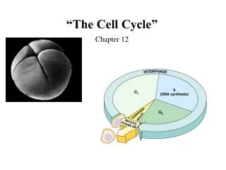
The Mitotic Cell Cycle Mitosis is just one part of the cell cycle. The mitotic (M) phase is usually the shortest part of the cell cycle. Mitotic cell division alternates with the much longer interphase, where the cell grows and copies its chromosomes. Interphase can be divided into subphases: -The G1 phase -The S phase (DNA synthesis) -The G2 phase During all three subphases, the cell grows by producing proteins and cytoplasmic organelles.
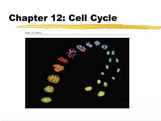
Mitosis is broken down into five subphases. • Prophase • Prometaphase • Metaphase • Anaphase • Telophase and Cytokinesis
Prophase: The chromatin fibers become more tightly coiled, condensing into discrete chromosomes. The nucleoli disappear, and each duplicated chromosome appears as two identical sister chromatids joined together. In the cytoplasm the mitotic spindle begins to form, which is made of microtubules extending from the two centrosomes. Prometaphase: The nuclear envelope fragments. The chromosomes have become more condensed, and the microtubules extend from each pole toward the middle of the cell. Each of the two chromatids of a chromosome has a specialized structure called a kinetochore, located at the centromere region. Microtubules attach to the kinetochores.
Metaphase: The centrosomes are now at opposite poles of the cell. The chromosomes line up on the metaphase plate with their centromeres on the plate. The entire apparatus of microtubules is called the spindle. Anaphase: The paired centromeres of each chromosome separate, separating the sister chromatids from each other. Each chromosome begins moving to the opposite pole of the cell as the kinetochore microtubules shorten. The poles of the cell move farther apart as the nonkinetochore microtubules lengthen.
Telophase and Cytokinesis: At telophase, the daughter nuclei form at the two poles of the cell. Nuclear envelopes arise from the fragments of the parent cell’s nuclear envelope and other portions of the endomembrane system. The chromatin fiber of each chromosome becomes less tightly coiled. Cytokinesis, the division of the cytoplasm, is usually well underway by this time. In animal cells, cytokinesis involves the formation of a cleavage furrow, which pinches the cell in two. Cell Plate
Mitosis in eukaryotes may have evolved from binary fission in bacteria • Prokaryotes reproduce by binary fission, literally meaning “division in half.”
Regulation of the Cell Cycle • The sequential events of the cell cycle are directed by the cell cycle control system, a cyclically operating set of molecules in the cell that both triggers and coordinates key events in the cell cycle. • A checkpoint in the cell cycle is a critical control point where stop and go-ahead signals can regulate the cycle. Animal cells have built in stop signals that halt the cell cycle until overridden by go-ahead signals. The signals report whether crucial cellular processes up to that point have been completed correctly and whether or not the cell should proceed. Three major checkpoints are found in the G1, G2, and M phases. • If a cell does not receive a go ahead-signal, it will exit the cycle, switching into a nondividing state called the G0 phase.
Cyclins and Cyclin-Dependent Kinases • Flunctuations in the abundance and activity of cell cycle control molecules pace the events of the cell cycle. Some of these molecules are protein kinases, enzymes that activate or inactivate other proteins by phosphorylating them. Protein kinases give the go-ahead signals at the G1 and G2 checkpoints. • The kinases that drive the cell cycle are always present, but are in the inactive form much of the time. To be active, a kinase has to be attached to a cyclin, which is a protein. These kinases are called cyclin-dependent kinases, or Cdks. • The activity of a Cdk rises and falls with changes in the concentration of its cyclin partner.
The first cyclin-Cdk complex discovered is called MPF, or “maturation-promoting factor” • The peaks of MPF activity correspond to the peaks of cyclin concentration. The cyclin level rises during interphase, then drops during mitosis. • MPF triggers the cell’s passage past the G2 checkpoint into M phase. It causes the nuclear envelope to fragment by phosphorylating proteins of the nuclear lamina. • Later in the M phase, MPF switches itself off by starting a process that leads to the destruction of its cyclin. • The non-cyclin part, Cdk, still exists in the cell, but is inactive until it associates with new cyclin molecules synthesized during interphase of the next round of the cycle.
Internal Signals: Messages from the Kinetochores • The M phase checkpoint will not allow anaphase to start unless all of the chromosomes are properly attached to the spindle at the metaphase plate. This ensures that the daughter cells do not end up with a missing or extra chromosome. • A signal that delays anaphase originates at kinetochores that are not yet attached to spindle microtubules. These proteins trigger a signaling pathway that keeps an anaphase-promoting complex (APC) in an inactive state. • When all the kinetochores are attached to the spindle, the “wait” signal stops. The APC then becomes active and triggers the breakdown of cyclin and the inactivation of proteins holding the sister chromatids together, allowing then to separate.
External Signals: Growth Factors • There are many external factors, both chemical and physical, that can influence cell division. • A growth factor is a protein released by certain body cells that stimulates other cells to divide. • Ex: platelet-derived growth factor (PDGF), which is made by blood cells called platelets. Fibroblasts, a type of connective cell tissue, have PGDF receptors on their plasma membranes. PGDF molecules bind to these receptors, which leads to stimulation of cell division. • When an injury occurs, platelets release PGDF, which causes fibroblasts to grow, healing the wound.
Density-dependent inhibition describes when crowded cells stop dividing. When a cell population reaches a certain density, the amount of required growth factors and nutrients available to each cell becomes insufficient to allow continued cell growth. • Most animal cells also exhibit anchorage dependence. In order to divide, they must be attached to something, such as the inside of a culture jar or the extracellular matrix of a tissue.
Cancer cells do not exhibit density-dependent inhibition or anchorage dependence. • They divide excessively and invade other tissues • Cancer cells can go on dividing indefinitely if they are given a continual supply of nutrients • Nearly all normal mammalian cells divide only about 20 to 50 times before they stop dividing. • Cancer starts in the body when a single cell undergoes transformation, the process that converts a normal cell to a cancer cell. The body’s immune system normally recognizes the cell and destroys it, but sometimes it evades destruction. • The cell can then divide to form a tumor, a mass of abnormal cells. It the cells remain at the original site, it is called a benign tumor, which mostly does not cause serious problems. • If the tumor becomes invasive enough to impair the functions of one or more organs, it is called a malignant tumor. • The spread of cancer cells to locations distant from the original site is called metastasis.