Download
1 / 70
720 likes | 793 Views
Explore the importance of cell division in life continuity and the evolution of mitosis from prokaryotes to eukaryotes, including the phases of the cell cycle and chromosome structure in eukaryotic cells. See how various organisms carry out cell division and its significance in tissue renewal, growth, and development.

E N D
Figure 12.1 • Overview: The Key Roles of Cell Division • The continuity of life: • is based upon the reproduction of cells, or cell division
100 µm (a) Reproduction. An amoeba, a single-celled eukaryote, is dividing into two cells. Each new cell will be an individual organism (LM). Figure 12.2 A Unicellular organisms: • Reproduce by cell division.
Origin of replication Cell wall Plasma Membrane E. coli cell Bacterial Chromosome Chromosome replication begins. Soon thereafter, one copy of the origin moves rapidly toward the other end of the cell. Two copies of origin 1 Replication continues. One copy ofthe origin is now at each end of the cell. 2 Origin Origin Replication finishes. The plasma membrane grows inward, and new cell wall is deposited. 3 Two daughter cells result. 4 • In binary fission: • The bacterial chromosome replicates • The two daughter chromosomes actively move apart Figure 12.11
Cell Replication in Bacteria • Cell division in bacteria is controlled by the size of the cell; volume of the cytoplasm. • Bacteria replicate by binary fission. • >22 enzymes copy the DNA as a circle. • Both copies of the DNA are attached to the plasma membrane.
The Evolution of Mitosis • Since prokaryotes preceded eukaryotes by billions of years: • It is likely that mitosis evolved from bacterial cell division • Certain protists: • Exhibit types of cell division that seem intermediate between binary fission and mitosis carried out by most eukaryotic cells
(a) Prokaryotes. During binary fission, the origins of the daughter chromosomes move to opposite ends of the cell. The mechanism is not fully understood, but proteins may anchor the daughter chromosomes to specific sites on the plasma membrane. Bacterial chromosome Chromosomes (b) Dinoflagellates. In unicellular protists called dinoflagellates, the nuclear envelope remains intact during cell division, and the chromosomes attach to the nuclear envelope. Microtubules pass through the nucleus inside cytoplasmic tunnels, reinforcing the spatial orientation of the nucleus, which then divides in a fission process reminiscent of bacterial division. Microtubules Intact nuclear envelope (c) Diatoms. In another group of unicellular protists, the diatoms, the nuclear envelope also remains intact during cell division. But in these organisms, the microtubules form a spindle within the nucleus. Microtubules separate the chromosomes, and the nucleus splits into two daughter nuclei. Kinetochore microtubules Intact nuclear envelope Kinetochore microtubules (d) Most eukaryotes. In most other eukaryotes, including plants and animals, the spindle forms outside the nucleus, and the nuclear envelope breaks down during mitosis. Microtubules separate the chromosomes, and the nuclear envelope then re-forms. Centrosome Fragments of nuclear envelope A hypothetical sequence for the evolution of mitosis Figure 12.12 A-D
200 µm 20 µm (c) Tissue renewal. These dividing bone marrow cells (arrow) will give rise to new blood cells (LM). (b) Growth and development. This micrograph shows a sand dollar embryo shortly after the fertilized egg divided, forming two cells (LM). Figure 12.2 B, C Multicellular organisms depend on cell division for: • Development from a fertilized cell • Growth • Repair
Cell Division in Eukaryotic Cells • Eukaryotic cells are much larger. • Also have larger genomes (the sum of an organisms’s genetic information). • Eukaryotic DNA is organized into much more complex structures. • Why?
Figure 12.3 50 µm The DNA molecules in a cell • Are packaged into chromosomes.
Chromosomes of Eukaryotes • Chromosomes are composed of chromatin - a DNA/protein complex. • Chromatin = 40% DNA + 60% Protein. • Every 200 nucleotides , the DNA duplex coils around a core of eight histone proteins = Nucleosome.
Regions of Chromosomes are Not the Same • Condensed portions of chromatin - Heterochromatin - are not being expressed; genes are turned off. May never be turned on. • Other portions of chromosome are decondensed and are being actively transcribed - Euchromatin. These areas are only condensed during cell division.
How Many Chromosomes are in Cells? • Most body or somatic cells have two copies of almost identical chromosomes - diploid. Ex. Humans have 23 types of chromosomes X 2 = 46 (2n or diploid number). • Sex cells or gametes (egg and sperm) have only one copy of each chromosome - haploid. Ex. Human sperm/egg = 23 chromosomes.
Continued • Some somatic cells are truly unusual. • Human liver cells have 4 copies of all chromosomes - tetraploid. • Other cells RBC’s have no nucleus and therefore, no chromosomes.
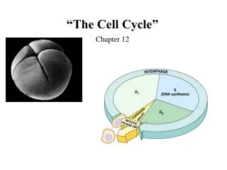
INTERPHASE S(DNA synthesis) G1 CytokinesisMitosis G2 MITOTIC(M) PHASE Figure 12.5 Phases of the Cell Cycle • The cell cycle consists of • The mitotic phase • Interphase G0
Chromosome Terminology • The two copies of each chromosome in somatic cells - homologous chromosomes (homologues). Are these the same? • Before cell division, each homologue replicates --> two sister chromatids that are joined at the centromere.
0.5 µm A eukaryotic cell has multiplechromosomes, one of which is represented here. Before duplication, each chromosomehas a single DNA molecule. Chromosomeduplication(including DNA synthesis) Once duplicated, a chromosomeconsists of two sister chromatidsconnected at the centromere. Eachchromatid contains a copy of the DNA molecule. Centromere Sisterchromatids Separation of sister chromatids Mechanical processes separate the sister chromatids into two chromosomes and distribute them to two daughter cells. Centromeres Sister chromatids Figure 12.4 • Each duplicated chromosome: • Has two sister chromatids, which separate during cell division
PROMETAPHASE G2 OF INTERPHASE PROPHASE Centrosomes(with centriole pairs) Aster Fragmentsof nuclearenvelope Early mitoticspindle Kinetochore Chromatin(duplicated) Centromere Nonkinetochoremicrotubules Kinetochore microtubule Nucleolus Chromosome, consistingof two sister chromatids Nuclearenvelope Plasmamembrane Figure 12.6 • Mitosis consists of five distinct phases • Prophase • Prometaphase
METAPHASE ANAPHASE TELOPHASE AND CYTOKINESIS Metaphaseplate Cleavagefurrow Nucleolusforming Nuclear envelopeforming Daughter chromosomes Centrosome at one spindle pole Spindle Figure 12.6 • Metaphase • Anaphase • Telophase
The Mitotic Spindle: A Closer Look • The mitotic spindle: • Is an apparatus of microtubules that controls chromosome movement during mitosis.
Aster Centrosome MetaphasePlate Sisterchromatids Kinetochores Overlappingnonkinetochoremicrotubules Kinetochores microtubules 0.5 µm Microtubules Chromosomes Figure 12.7 Centrosome 1 µm • Some spindle microtubules: • Attach to the kinetochores of chromosomes and move the chromosomes to the metaphase plate
EXPERIMENT 1 The microtubules of a cell in early anaphase were labeled with a fluorescent dye that glows in the microscope (yellow). Kinetochore Spindlepole Figure 12.8 • In anaphase, sister chromatids separate: • And move along the kinetochore microtubules toward opposite ends of the cell
Nonkinetechore microtubules from opposite poles • Overlap and push against each other, elongating the cell • In telophase: • Genetically identical daughter nuclei form at opposite ends of the cell
Cell Cycle G0
Cleavage furrow 100 µm Contractile ring of microfilaments Daughter cells Figure 12.9 A (a) Cleavage of an animal cell (SEM) Cytokinesis: A Closer Look • In animal cells • Cytokinesis occurs by a process known as cleavage, forming a cleavage furrow
Vesiclesforming cell plate 1 µm Wall of patent cell Cell plate New cell wall Daughter cells Figure 12.9 B (b) Cell plate formation in a plant cell (SEM) • In plant cells, during cytokinesis: • A cell plate forms.
Plant Cells Lack centrioles No aster Assemble membrane components in the interior- cell plate Deposts cellulose- new cell wall. Animal Cells Use centrioles Centrioles develop an aster Actin filaments constrict the cell -cleavage furrow. Plant vs. Animal Mitosis
Cell Cycle Control • Internal clocks are not flexible. • Eukaryotic cells use Check Points- enzymes that survey conditions of the cell and act as “go/no go” switches. • Regulated by feedback from the cell.
G1 checkpoint Control system S G1 G2 M M checkpoint Figure 12.14 G2 checkpoint The Cell Cycle Control System • The sequential events of the cell cycle • Are directed by a distinct cell cycle control system, which is similar to a clock
G0 G1 checkpoint G1 G1 (a) If a cell receives a go-ahead signal at the G1 checkpoint, the cell continues on in the cell cycle. (b) If a cell does not receive a go-ahead signal at the G1checkpoint, the cell exits the cell cycle and goes into G0, a nondividing state. Figure 12.15 A, B • The clock has specific checkpoints: • Where the cell cycle stops until a go-ahead signal is received
Three Principal Checkpoints • G1 Checkpoint (also called START)- end of G1/beginning of S- decision to replicate DNA. • Surveys cell size and environmental conditions.
G1 Checkpoint Replicate DNA Cell Size? Yes Favorable Conditions? No Grow or GO