Download
1 / 1
20 likes | 172 Views
Autoantibody Reactivity to Tumor Associated Antigens as a Biomarker for Early Lung Cancer. J Hung 1 , M Jagen 1 , AK Greenberg 1 , E Tan 2 , D Naidich 1 , H Steck 3 , G Fung 3 , M Salganikoff 3 , B Rao 3 , BK Phalan 1 , E Eylers 1 and WN Rom 1

E N D
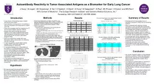
Autoantibody Reactivity to Tumor Associated Antigens as a Biomarker for Early Lung Cancer J Hung1, M Jagen1, AK Greenberg1, E Tan2, D Naidich1, H Steck3, G Fung3, M Salganikoff 3, B Rao3, BK Phalan1, E Eylers1 and WN Rom1 NYU School of Medicine1, The Scripps Research Institute2 and Siemens Medical Solutions, Inc.3 Funded by: NIH U01CA86137, M01RR-00096 Methods Introduction Summary of Results Results Percent of Lung Cancer Cases and Smoking Controls with positive ELISA (defined as > 2x [NHC mean + 3 std dev]) Patient Demographics • Lung cancer is the leading cause of death among all cancer types. • Early detection of lung cancer in its earliest or even pre-neoplastic form may lead to a higher cure rate. • Some preneoplasias may be visualized on CT scan as ground glass opacities (GGOs). • Studies have demonstrated that cancer patients’ sera contain antibodies that reacts with autologous intracellular proteins.These proteins are known as tumor associated antigens (TAA) • Sera from individuals enrolled in the NYU Lung Cancer Biomarker Center (NYU LCBC) of NCI Early Detection Research Network were analyzed for reactivity to tumor associated antigens (TAAs) • Patient population: • Individuals were categorized in the following groups • Non-smoking healthy control (NHC), n=40. • Lung cancer patients, n=17. • Smokers with solid nodules (SN) seen on initial screening chest CT, n=49. • Smokers with GGOs seen on initial screening chest CT, n=47. • Smokers with normal CT (Smoking controls), n=45. • The cancer patients and smokers with GGOs were consecutive cases enrolled in the NYU LCBC. Smoking controls and individuals with stable solid noduels were matched for age and smoking history to the cancer and GGO patients (combined). • Smokers were defined as individuals with > 20 pack years of cigarettes smoking history • Smoking controls had a higher level of reactivity to a panel of TAAs than non-smoking controls. • When compared with smoking controls, reactivity to c-myc and p62 was significantly elevated in both patients with lung cancer and in individuals with possible preneoplastic lesions (GGOs). Both the mean levels of reactivity were increased, and the number of individuals with reactivity above a cut-off value (>2x mean normal +3SD) were increased. • In individuals with GGOs, significantly more people also had increased reactivity to cyclin B1 and decreased reactivity to cyclin D1 compared with smoking controls. • Individuals with stable solid nodules (presumably benign) also had increased reactivity to c-myc compared with smoking controls * p-value <0.05 when compared to Smokers with normal chest CT Mean ELISA Results in Optical Density Percent of Smokers with GGOs and Smoking Controls with positive ELISA (defined as > 2x [NHC mean + 3 std dev]) • GGOs are defined as nodules with hazy attenuation without obscuration of underlying vascular markings. • In high risk patients, ground-glass opacity nodules (GGO) and solid nodules are commonly seen on Chest CTs. • In one study (Nakata et al., Chest 2002) all the GGOs ≤ 2 cm that persisted for 3 months or more were malignant or premalignant lesions. Of 43 persistent focal GGOs, 53% were bronchioalveolar carcinoma (BAC), 26% were adenocarcinoma and 21% were preneoplasias (atypical adenomatous hyperplasia—AAH). • In this study, we determined level of autoantibody to a panel of TAAs in individuals with lung cancer, GGOs, solid nodules and normal CTs. Ground Glass Nodule Conclusion *p-value <0.05 when compared to non-smoking healthy control • Our results suggest a pattern of autoantibody reactivity to a panel of TAAs may distinguish patients with lung cancer and preneoplastic lesions from smokers with normal CTs or benign lesions. • The finding of a similar pattern of reactivity in both patients with lung cancer and in individuals with possible preneoplastic lesions, suggests that this assay may prove useful for the identification of these very early stages of lung cancer. • Further studies, with increased numbers and perhaps additional TAAs, are needed to validate these results and increase the sensitivity and specificity of the assay. Percent of Smokers with SNs and Smoking Controls with positive ELISA (defined as > 2x [NHC mean + 3 std dev]) • Enzyme-linked immunosorbent assay (ELISA) to TAAs were performed on sera • Tumor associated antigens (TAA) • P53 • c-myc • Insulin-like growth factor 2, binding protein 1 (IMP1) • p62/IMP2 • IMP3/Koc • CyclinA • Cyclin B1 • Cyclin D1 • CDK2 • Survivin • Optical density (OD) value at 490 nm were recorded • The cut-off value for positive ELISA was defined as having >2 times of mean OD value of non-smoking normal + 3 standard deviation. • Statistical analysis was performed using SPSS software O.D. Hypothesis • We hypothesized that autoantibody reactivity level to a panel TAAs would be elevated in the serum of patients with lung cancer and preneoplasia (represented by persistent GGOs on CT); and that this reactivity to TAAs could be a biomarker for early lung cancer detection. O.D.