Bacterial Morphology and Structure
Bacterial Morphology and Structure Xiao-Kui Guo PhD http://basic.shsmu.edu.cn/passw/micro2/index.asp SIZE OF BACTERIA Unit for measurement : Micron or micrometer,μm: 1μm=10-3mm Size: Varies with kinds of bacteria, and also related to their age and external environment.

Bacterial Morphology and Structure
E N D
Presentation Transcript
Bacterial Morphology and Structure Xiao-Kui Guo PhD http://basic.shsmu.edu.cn/passw/micro2/index.asp
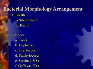
SIZE OF BACTERIA • Unit for measurement : Micron or micrometer,μm: 1μm=10-3mm • Size: Varies with kinds of bacteria, and also related to their age and external environment. • Cocci: sphere, 1μm • Bacilli: rods , 0.5-1 μm in width -3 μm in length • Spiral bacteria: 1~3 μm in length and 0.3-0.6 μm in width
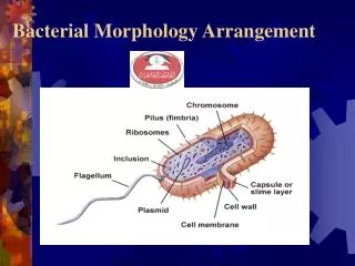
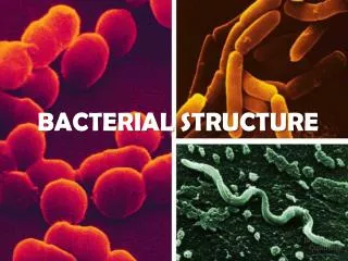
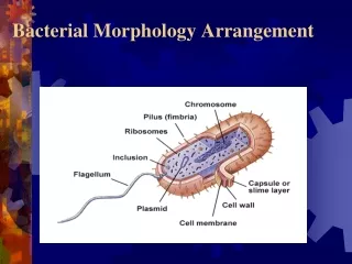
Structure of Bacteria Essential structures cell wall 细胞壁cell membrane 细胞膜Cytoplasm 细胞质nuclear material核质 Particular structures capsule 荚膜 flagella 鞭毛 pili 菌毛 spore芽胞
Flagellum Nucleoid Cell membrane Cell wall Gram + Pili Gram - Granule Capsule Cell (inner) membrane Outer membrane Ribosomes Cell wall 1884: Christian Gram: First publication for the Gram stain method) Editor's note: I would like to testify that I have found the Gram method to be one of the best and for many cases the best method which I have ever used for staining Schizomycetes. Gram, C. 1884. Ueber die isolirte Farbung der Schizomyceten in SchnittÄund Trockenpraparaten. Fortschritte der Medicin, Vol. 2, pages 185-189.
Cell wall • Situation: outmost portion. 15-30nm in thickness, 10%-25% of dry weight.
Cell wall :Common peptidoglycan layer • A backbone of N-acetyl glucosamine and N-acetylmuramic acid: Both discovered in Gram positive and Gram negative bacteria. • A set of identical tetrapeptide side chain attached to N-acetyl-muramic acid: different components and binding modes in Gram positive and Gram negative bacteria. • A set of identical peptide cross bridges: only in Gram positive bacteria
Special components of Gram positive cell wall Teichoic acid SPA / M POTEIN
Functions of Cell Wall • Maintaining the cell's characteristic shape- the rigid wall compensates for the flexibility of the phospholipid membrane and keeps the cell from assuming a spherical shape • Countering the effects of osmotic pressure • Providing attachment sites for bacteriophages • Providing a rigid platform for surface appendages- flagella, fimbriae, and pili all emanate from the wall and extend beyond it • Play an essential role in cell division • Be the sites of major antigenic determinantsof the cell surface。 • Resistance of Antibiotics
Wall-less forms of Bacteria. • When bacteria are treated with 1) enzymes that are lytic for the cell wall e.g. lysozyme or 2) antibiotics that interfere with biosynthesis of peptidoglycan, wall-less bacteria are often produced. • Usually these treatments generate non-viable organisms. Wall-less bacteria that can not replicate are referred to as spheroplasts (when an outer membrane is present) or protoplasts (if an outer membrane is not present). • Occasionally wall-less bacteria that can replicate are generated by these treatments (L forms).
Cell membrane • Site of biosynthesis of DNA, cell wall polymers and membrane lipids.Selective permeability and transport of solutes into cells • Electron transport and oxidative phosphorylation • Excretion of hydrolytic exoenzymes
Mesosomes • Mesosomes are specialized structures formed by convoluted inveigh-nations of cytoplasmic membrane, and divided into septal and lateral mesosome.
Cytoplasm • Composed largely of water, together with proteins, nucleic acid, lipids and small amount of sugars and salts • Ribosomes: numerous, 15-20nm in diameter with 70S; distributed throughout the cytoplasm; sensitive to streptomycin and erythromycin site of protein synthesis • Plasmids: extrachromosomal genetic elements • Inclusions: sources of stored energy, e,g volutin
Plasmids are small,circular/line,extrachromosomal,double-stranded DNA molecules。They are capable of self-replication and contain genes that confer some properties,such as antibiotic resistance,virulence factors。Plasmids are not essential for cellular survival. Plasmid Inclusions of Bacteria • Inclusions are aggregates of various compounds that are normally involved in storing energy reserves or building blocks for the cell. Inclusions accumilate when a cell is grown in the presence of excess nutrients and they are often observed under laboratory conditions. granulose
Nucleus • Lacking nuclear membrane, absence of nucleoli, hence known as nucleic material or nucleoid, one to several per bacterium.
Attachment • Protection from phagocytic engulfment. • Resistance to drying. • Depot for waste products. • Reservoir for certain nutrients. • protection Capsules and slime layers • These are structures surrounding the outside of the cell envelope. They usually consist of polysaccharide; however, in certain bacilli they are composed of a polypeptide (polyglutamic acid). They are not essential to cell viability and some strains within a species will produce a capsule, whilst others do not. Capsules are often lost during in vitro culture.
Some bacterial species are mobile and possess locomotory organelles - flagella. Flagella consist of a number of proteins including flagellin • The diameter of a flagellum is thin, 20 nm, and long with some having a length 10 times the diameter of cell. Due to their small diameter, flagella cannot be seen in the light microscope unless a special stain is applied. Bacteria can have one or more flagella arranged in clumps or spread all over the cell. Flagella Monotrichate/Amphitrichate/Lophotrichate/Peritrichate • Identification of Bacteria • Pathogenesis • Motility of bacteria
Pili • Pili are hair-like projections of the cell , They are known to be receptors for certain bacterial viruses. Chemical nature is pilin • Classification and Function • Common pili or fimbriae: fine , rigid numerous, related to bacterial adhesion • Sex pili: longer and coarser, only 1-4, related to bacterial conjugation
Endospores (spores) • Identification of Bacteria • Pathogenesis • Resistance • Dormant cell • Resistant to adverse conditions • - high temperatures • - organic solvents • Produced when starved • Contain calcium dipicolinate • DPA, Dipicolinic acid • Bacillus and Clostridium
Methods Microscope • Light Microscope • Electron Microscope • Darkfield Microscope • Phase Contrast Microscope • Fluorescence Microscope • Cofocal Microscope) Staining Methods • Simple staining; • Differential staining ( Gram stain, Acid-fast stain), • Special staining( Negative stain, Spore stain, Flagella stain)