BACTERIAL STRUCTURE
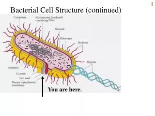
BACTERIAL STRUCTURE. Bacterial cell ( coccus or bacillus) will have some structures common Cell wall Cell membrane Cytoplasm Ribosomes Chromosome I ntra-cellular structures Plasmid Inclusion bodies Extra-cellular structures Capsule Fimbriae Flagella . CELL WALL

BACTERIAL STRUCTURE
E N D
Presentation Transcript
Bacterial cell (coccus or bacillus) will have some structures common • Cell wall • Cell membrane • Cytoplasm • Ribosomes • Chromosome • Intra-cellular structures • Plasmid • Inclusion bodies • Extra-cellular structures • Capsule • Fimbriae • Flagella
CELL WALL It is a layers of cell envelope lying between the cytoplasmic membrane and the capsule Gram positive bacteria cell wall mainly consists of peptidoglycan and teichoic acid Gram negative bacteria includes peptidoglycan, lipoprotein, outer membrane and lipopolysaccharidelayers
Bacteria have a complex cell wall consisting of peptidoglycan (also called murein, mucopeptide) This complex polymer consists of three parts: • A backbone consisting of alternating units of NAG (N acetylglucosamine) and NAM (N-acetylmuramic acid). • Tetrapeptideside chain attached to NAM • Peptide cross-bridges, which are short chains of amino acids that crosslink the backbone.
GRAM POSITIVE BACTERIAL CELL WALL • Contain 40 sheets of peptidoglycancomprising up to 50% of cell wall material • Peptidoglycanof Gram positive cells to be 20-80 nm thick • Contain additional substances such as teichoic acid and teichuronic Acid • These are water soluble polymers of ribitol or glycerol • There are two types of teichoicacid: • Wall teichoicacid (linked to peptidoglycan) • Lipoteichoicacid (linked to membrane)
Some gram positive bacteria may lack wall teichoicacid but all contain lipoteichoicacid • The teichoic acid constitutes major antigens of cells • Teichoicacid binds to Magnesium ions and plays a role in supply of this ion to the cell • On digestion of peptidoglycan from the cell, gram positive cells lose their cell walls and become protoplasts • Gram negative cells become spheroplasts
GRAM NEGATIVE BACTERIAL CELL WALL • Gram negative cells have thin layer of peptidoglycan (approximately 10 nm) • There appears to be only one or two sheets of peptidoglycan, comprising 5-10% of cell wall • They have an additional outer membrane acting as permeability barrier • The space between the inner and outer membranes is known as the periplasmicspace, which contains digestive enzymes and other transport proteins
Gram negative cell walls contain three components that lie outside the peptidoglycanlayer: • Lipoprotein:Stabilizes the outer membrane by anchoring it to peptidoglycan • Outer membrane is phospholipidbilayer in which the outer phospholipids are replaced by lipopolysaccharides • Contain several important porins, which specifically allow transport of solutes. • Lipopolysaccharide
Lipopolysaccharide • Polysaccharide core • Complex lipid called Lipid A • Terminal series of repeat units • The polysaccharide core is similar in all gram negative bacteria • Each species contains unique terminal repeat units • LPS is toxic in nature and is called endotoxin • It is firmly bound to the cell wall and released only when cell is lysed • Endotoxincan trigger fever and septic shock in gram negative infections
LPS confers a negative charge and also repels hydrophobic molecules such as bile in the intestine • LPS is split into Lipid A and polysaccharide, all the toxicity is associated with Lipid A and polysaccharide • Lipid A represents the major surface antigen of bacterial cell • This antigen is designated as somatic “O” antigen and is used in serological typing of species • Antigenic specificity is conferred by the terminal repeat units
Bacteria are divided into two groups based upon the composition of their cell walls. • Gram positive : two layers ( lipid, peptidoglycan – sugar/amino acids network) • Gram negative : three layers, lipid, peptidoglycan, and lipopolysaccharide Gram + Gram -
Significance of cell wall • Maintains cell shape, any cell that loses its cell wall, loses its shape • Protects bacteria from osmotic lysis • Acts as a barrier, protects cell contents from external environment • Determines reactivity to Gram stain, cells become gram negative if they lose cell wall • Attachment site for flagella • Site of action of certain antimicrobial agents (E.g. Penicillins,Cephalosporins) • Confer specific antigenicity to a strain/species that can be exploited to detect and identify an isolate
Substances acting against cell wall Lysozyme, an enzyme found in tears and saliva breaks down a component of cell walls Antibiotics that inhibits cell wall synthesis such as Penicillins and cephalosporins Autolytic enzymes produced by some bacteria such as Streptococcus pneumoniae
CELL MEMBRANE • Cell membrane or cytoplasmic membrane is a typical unit membrane • Composed of phospholipids (40%) and proteins (60%) • It measures approximately 5-10 nm in thickness • It lies below the peptidoglycan layer of the cell wall and encloses the cytoplasm • The arrangement of proteins and lipids to form a membrane is called the Fluid Mosaic Model
Specialized structures called mesosomes or chondroids are formed from the convoluted invaginations of cytoplasmic membrane. • Mesosomes are of two types: • Septalmesosome • Lateral mesosome. • The bacterial chromosome is attached to the septalmesosome • During cell division, the septalmesosome participates in theformation of cross-walls • Mesosomesare more prominant in gram positive bacteria.
Functions of cell membrane • Selectively permeable barrier: substances are limited by pore sizes and the hydrophobic nature of the membrane • Integral (transmembrane) proteins form channels and act as carriers • Peripheral proteins can act as receptors and as enzymes for metabolic reactions • Electron transport and oxidative phosphorylation are located in the cell membrane • Excretion of hydrolytic enzymes • Site of initiation of cell wall synthesis • Site of synthesis of phospholipids • Bear receptors and proteins of sensory transduction system
CYTOPLASM It is the portion of the cell that lies within the cytoplasmicmembrane It is gel-like in consistency and includes the procaryotic chromosome and ribosomes The cytoplasm does not exhibit any internal mobility (cytoplasmic streaming) The cytoplasm also lacks organelles such as mitochondria, golgi apparatus or endoplasmic reticulum Cytoplasm stains uniformly in young cultures. Recent studies suggest that some bacteria (Bacillus subtilis) possess cytoskeleton.
Constituents of cytoplasm include: • Proteins (including enzymes) • Vitamins • Ions • Nucleic acids and their precursors • Amino acids and their precursors • Carbohydrates and their derivatives • Fatty acids and their derivatives.
Chromosome • The chromosome in bacteria is typically a single, closed circle DNA • It is concentrated in a nucleoidregion • It is not membrane bound as in eukaryotes • Some bacteria possess smaller extrachromosomal pieces of DNA called plasmids • Plasmids replicate independently of the chromosome • Plasmid carry genes that are not essential for cell • survival
Chromosome • The chromosome is attached to an invagination of the cytoplasmicmembrane called Mesosome • Mitotic apparatus and nuclear membrane are completely lacking • The length of E.coli chromosome is approximately 1.4 mm but is condensed inside the cell by supercoiling • DNA is negatively charged hence bind readily to basic dyes • It can be demonstrated by Feulgen stain or by electron microscopy
Ribosomes • Bacterial cells contain thousands of ribosomes, which are the sites of protein synthesis • The distinct granular appearance of procaryotic cytoplasm is due to the presence and distribution of ribosomes • They aggregate to form structures known as polysomes • Bacterial ribosomes are termed 70 S (Svedberg units)
Inclusion bodies • Intra-cytoplasmicinclusions can be vacuoles, crystals or storage bodies • Bacteria often store reserve material in the form of insoluble cytoplasmicgranules • Inclusions accumulate when a cell is grown in the presence of excess nutrients
Inclusion bodies Various examples of these bodies are: Starch/Glycogen granules - blue-greens and enteric bacteria Poly-ß-hydroxybutyrate granules - Azotobacter and Rhizobium Nitrogen-reserve granules - blue-greens Sulphur inclusions – Thiotrix Lipid inclusions Volutin granules – on staining with Sudan Black Dye
FLAGELLA • Some bacteria are motile and almost all motile bacteria possess flagella as the organ of locomotion • Such bacteria tend to move towards or away from the source of stimulus • These stimuli can be chemicals (chemotaxis), light (phototaxis), air (aerotaxis) or magnetism (magnetotaxis)
Structure • Procaryoticflagella are much thinner than eukaryotic flagella • They lack the typical 9 + 2 arrangement of microtubules • They are approximately 3-20μm long and end in a square tip • The bacterial flagellum is a non contractile, composed of single kind of protein subunit called flagellin • It is anchored to the bacterial cytoplasmic membrane and cell well
A flagellum comprises of three parts: • Filament • Hook • Basal body • The flagellum is attached to the cell body by hook and basal body • Hook and basal body are embedded in the cell envelope, the filament is free • If a flagellum is cut off it will regenerate until reaches a maximum length • This is so because the growth is not from base, but from tip
The basal body bears a set of rings: • One pair in gram positive bacteria • Two pairs in gram negative bacteria • Rings named S and M are common to both, the rings names P and L are found only in gram negative bacteria • Rings in the basal body rotate relative to each other causing the flagella to turn like a propeller • The energy to drive the basal body is obtained from the proton motive force • Bacteria move at average speed of 50μm/sec, the fastest being Vibriocholerae that moves 200μm/sec.
Flagella arrangements are: • Monotrichous- a single flagellum at one pole (also called polar flagellum) E.g. Vibriocholerae • Amphitrichous- single flagellum at both poles. Eg. Spirilla • Lophotrichous- two or more flagella at one or both poles of the cell E.g. Spirillumundula • Peritrichous- completely surrounded by flagella E.g. E.coli
Other mechanisms of bacterial locomotion include gliding and motion by axial filament contraction • Gliding is movement of bacteria along solid surfaces by an unknown mechanism • Spirochetes have internally-located axial filaments or endoflagella • Axial filaments wrap around the spirochete towards the middle from both ends • They are located above the peptidoglycan cell wall but below the outer membrane
Significance of flagella • Primarily function is motility (chemotaxis, aerotaxis, phototaxisetc) • Positive taxis is movement toward a favorable environment whereas negative taxis is movement away from a repellent • Flagella can help in identifying certain types of bacteria. For example, Proteus species show ‘swarming’ type of growth on solid media. • Flagellar antigens are used to distinguish different species and strains of bacteria (serovars). Variations in the flagellar H antigen are used in serotyping.
FIMBRIAE AND PILI • Fimbriaeare short, hair-like structures made up of protein PILIN,present in many gram negative bacteria • Pili areanchored in the membrane and protrude through the cell wall to the outside of the cell • Fimbriaeare shorter and straighter than flagella and are more numerous • They are 0.5μm long and 10 nm thick • Since they are made up of protein, they are antigenic
The term piliis used to denote sex pili Sex pili acts to join bacterial cells for transfer of DNA from one cell to another by a process called conjugation. Significance: • They act as adhesins and allow bacteria to colonize cells. For example, Neisseria gonorrhoea • Fimbriaecan also detect chemical signals and are important in bacterial cell communication and biofilmformation. • Fimbriaealso act as receptors for bacteriophages • Fimbriaeof Streptococcus pyogenesare coated with M protein, which acts as an important virulence factor by adhering to host cells and resisting phagocytosis
L-FORMS, PROTOPLAST AND SPHEROPLASTS • When bacteria are treated with enzymes that hydrolyze the cell wall (e.g. lysozyme) or antibiotics that interfere with biosynthesis of peptidoglycan (penicillin), wall-less bacteria are often produced. • It liberates protoplastsfrom gram positive bacteria and spheroplastsfrom gram negative bacteria • Spheroplastsretain the outer membrane • These treatments generate wall-less, non-viable organisms that do not multiply • But if such cells can grow and divide, they are called L forms
INVOLUTION FORMS AND PLEOMORPHISM • Certain species of bacteria exhibit variation in shape and size of individual cells • This variation is known as pleomorphism • Swollen and aberrant forms are seen in ageing cultures of Nesseriagonorrhoeaeand Yersiniapestisor in the presence of high salt concentration • Such forms are known as involution forms • Both formsare believed to be the result of defective cell wall synthesis or due to the action of autolytic enzymes that digest their own cell wall.
Glycocalyx/Capsule/Slime • Gelatinous polysaccharide or polypeptide outer covering of certain bacteria is called glycocalyx • surround outside the cell envelope • Glycocalyxis referred to as a CAPSULEif it is firmly • attached to the cell wall • As a slime layer if loosely attached • The chemical nature of bacterial capsules are polysaccharides • These polymers are composed of repeating oligosaccharide units
Capsules may be weakly antigenic to strongly antigenic, depending on their chemical complexity • Capsules may be covalently linked to the underlying cell wall or just loosely bound to it • Bacteria with capsules form smooth (S) colonies while those without capsules form rough (R) colonies • Species may undergo a phenomenon called S-R variation whereby the cell loses the ability to form a capsule • Capsules are sometimes referred as K antigens (in Enterobacteriaceae) or as Vi antigen (in Salmonella typhi)
Significance • Capsules of pathogenic bacteria inhibit ingestion and killing by phagocytes • Prevent complement-mediated bacterial cell lysis • Protect the cells from lysozyme • Permit bacteria to adhere to cell surfaces leading to colonization and disease • Capsules can be a source of nutrients and energy to microbes • Streptococcus mutans, which colonizes teeth, ferments the sugar in the capsule and acid byproducts contribute to tooth decay
Prevent cell from drying out (desiccation) • Toxicity to the host cell • Capsules may protect cells from bacteriophages • Capsules play a role in antigenic mosaic • Capsules may trap ions • Examples: • Streptococcus pneumoniae, Streptococcus mutans, Klebsiellapneumoniae, Bacillus anthracis, Neisseria • meningitidis
SPORE • Under adverse conditions some bacteria such as Bacillus and Clostridium produce resistant survival forms termed endospores • This process is known as sporulation • Spores are resistant to extreme environmental conditions such as high temperatures, dryness, toxic chemicals (disinfectants, antibiotics), and UV radiation • On sporulation, the vegetative portion of the bacterium is degraded and the dormant endospore is released • The endospore then germinates, producing a single vegetative bacterium
Mechanism of sporulation DNA replicates and the cell divides asymmetrically A cytoplasmic membrane septum forms at one end of the cell A second layer of cytoplasmic membrane then forms around one of the DNA molecules to form a forespore Both of these membrane layers then synthesize peptidoglycanin the space between them to form the cortex Calcium dipocolinate is also incorporated into the forming endospore
Mechanism of sporulation Spore coat composed of a keratin-like protein then forms around the cortex Sometimes an outer membrane composed of lipid and protein and called an exosporium is also formed Finally, the remainder of the bacterium is degraded and the endospore is released There is no metabolic activity until the spore is ready to germinate Single vegetative cell gives rise to a single spore Sporulationgenerally takes around 15 hours.
Germination Favorablegrowth conditions signal the process of endospore germination Germination of a spore results in a break in the spore wall and the outgrowing of a new vegetative cell The newly formed vegetative cell is capable of growthand reproduction A single spore upon germination forms a single vegetative cell.
Germination occurs in following steps: 1. Activation: Even in the presence of favorable conditions, the spore will not germinate until its protective spore coat is not damaged Conditions such as heat, acidity, abrasion or compounds containing free sulphydrylgroups activate the spore to germinate
2. Initiation:On activation, the spore will germinate provided the environment is suitable Binding of effector stimulates autolytic enzymes that degrade the peptidoglycanof cortex Water is absorbed and calcium dipicolinate is released 3. Outgrowth: Once the cortex and outer layers is degraded, a new vegetative cell consisting of spore protoplast and its wall emerges This is followed by active biosynthetic activity and process terminates with cell division.
The resistance of endospores is due to a variety of factors: o Calcium-dipicolinate, abundant within the endospore, may stabilize and protect the endospore's DNA o Specialized DNA-binding proteins saturate the endospore'sDNA and protect it from heat, drying, chemicals, and radiation o The cortex may osmotically remove water from the endosporeand impart resistance to heat and radiation o DNA repair enzymes contained within the endosporeare able to repair damaged DNA during germination.