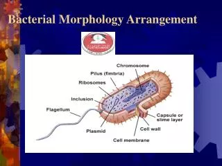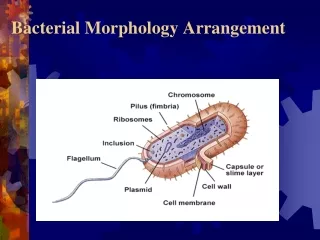
Bacterial Morphology Arrangement
380 likes | 991 Views
Discover the diverse shapes, sizes, and arrangements of bacteria, including coccus, bacillus, spirillum, vibrio, and pleomorphic forms. Learn about bacterial cell structure, appendages like flagella and pili, and unique features such as glycocalyx and cell walls. Gain insights into the functions and classifications based on bacterial morphology.

Bacterial Morphology Arrangement
E N D
Presentation Transcript
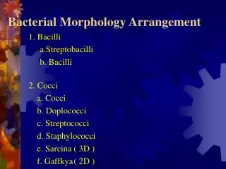
Bacterial Shapes, Arrangements, and Sizes Variety in shape, size, and arrangement but typically described by one of three basic shapes: coccus - spherical bacillus – rod coccobacillus – very short and plump ( Brucella abortus) Streptobacilli ( Bacillus subtilus) diplobacilli spirillum - helical, comma, twisted rod, spirochete – spring-like- flexible ( Treponema pallidum) vibrio – gently curved ( Vibrio cholera) Spirilla- rigid ( Borrelia species) Pleomorphic : variable in shape ( Corynebacterium) 2
Bacterial Cell Structure • Appendages - flagella, pili or fimbriae • Surface layers - capsule, cell wall, cell membrane • Cytoplasm - nuclear material, ribosome, mesosome, inclusions etc. • Special structure - endospore
Appendages 1. flagella Some rods and spiral form have this. a). function: motility b). origin : cell membrane flagella attach to the cell by hook and basal body which consists of set(s) of rings and rods Gram - : 2 sets of ring and rods, L, P, S, M rings and rods . e.g. E. coli Gram + : S, M rings and rods .e.g. B. megaterium
Flagella • Motility - movement • Swarming occurs with some bacteria • Spread across Petri Dish • Proteus species most evident • Arrangement basis for classification • Monotrichous; 1 flagella • Lophotrichous; tuft at one end • Amphitrichous; both ends • Peritrichous; all around bacteria
The structure of the bacterial flagella allows it to spin like a propeller and thereby propel the bacterial cell; clockwise or counter clockwise wave like motion. • Bacterial flagella provides the bacterium with mechanism for swimming toward or away from chemical stimuli, a behavior is knows as CHEMOTAXIX, chemosenors in the cell envelope can detect certain chemicals and signal the flagella to respond. - structure protein in nature: subunit flagellin ( globular protein)
2. Fimbriae and Pili Fimbriae: Shorter than flagella and straighter , smaller, hairlike appendages . Only on some gram- bacteria. a). function: adhere. Not involve in motility. One of the invasive mechanism on bacteria. Some pathogens cause diseases due to this (Antigenic characteristic). Prevent phagocytosis.
pili - sex factor. If they make pili, they are + or donors of F factor. It is necessary for bacterial conjugation resulting in the transfer of DNA from one cell to another. It have been implicated in the ability of bacteria to recognize specific receptor sites on the host cell membrane.
. Origin: Cell membrane . Position: common pili , numerous over the cell, usually called sex pile, 1-4/cell . Structure: composed of proteins which can be dissociated into smaller unit Pilin . It belongs to a class of protein Lectin which bond to cell surface polysaccharide.
Axial Filaments • Present in spirochetes ( Treponema pallidum cause syphilis) • Function is motility – gliding motility • Bundles of fibrils that arise at the ends of the cell
Axial filament Structurally similar to flagella Unique location under an outer membrane Spirochetes
Glycocalyx • Agelatinous polysaccharide and/or polypeptide outer covering. The glycocalyx can be identified by negative staining techniques. • Two types: • Slime layer - loosely organized and attached unorganized material that is easily removed. • Capsule - highly organized, tightly attached The layer is well organized and not easily washed off. • B. anthracis has a capsule of poly-D-glutamic acid, while S. pyogenes made of Hyaluronic acid.
. Glycocalyx: Capsule or slime layer It consists of polypeptide and polysaccharide on bacilli. Most of them have only polysaccharide. It is a protective layer that resists host phagocytosis. Medically important ( Streptococcus pneumonia).
Capsule and Slime layer play arole in Attachment and formation of biofilms
Capsules can serve numerous functions including -Virulence factors, protecting bacteria from phagocytosis by immune cells. Pathogens such as Streptococcus pneumoniae can cause pneumonia if protected by a capsule. -Permit bacteria to adhere to cell surfacesand structures such as medical implants, catheters and so on. This is an important first step in colonization and sometimes leads to disease. -Capsules can be a source of nutrients and energy to microbes. Streptococcus mutans, which colonizes teeth, ferments the sugar in the capsule and acid byproducts contribute to tooth decay. -Prevent cell from drying out (desiccation).
Bacterial cell wall The cell wall is the outer most component common to all bacteria( except Mycoplasma species which are bounded by cell membrane) some bacteria have surface feature external to the cell wall, such as a capsule, flagella and pilli.
Functions of the cell wall - maintains cell shape . - Acts as a barrier, protects cell contents from external environment . - maintains cell integrity/osmotic pressure in a hypotonic environment. - Determines reactivity to Gram stain. - Attachment site for flagella. -Contributes to sensitivity to certain antimicrobial agents and the immune system (antibodies, phagocytes).
Gram positive cell walls • Consist of a relatively thick layer of exposed peptidoglycan (60-90% of the cell wall) .Also called murein or mucopeptide
Gram positive cell walls -The peptidoglycan backbone consists of alternating units of 2 sugars called NAG (N-acetylglucosamine) and NAM (N-acetylmuramic acid) -The sugar backbone is crosslinked by short chains of amino acids. There are also side chains of tetrapeptides attached to NAM.
peptidoglycanlayercontributes to sensitivity to certain antimicrobial agents • The site of action of lysozyme is to break the bond that links G to M. The site of action of penicillin is to prevent the formation of the interpeptide bond that occasionally joins the peptide chains to one another. In either case, the result is the lysis of the bacterial cell.
Gram positive cell walls • cells stain purple due to retention of the crystal violet dye during the gram stain procedure. • Antigens called teichoic acids project out of the cell wall and aid in typing different gram positive bacteria. It is a polymerof glycerol or ribitol joined by phosphate groups , and cause septic shock.
Function of Teichoic acids: * Antigenic determinant * Participate in the supply of Mg to the cell by binding Mg++ * regulate normal cell division.