Download
1 / 18
190 likes | 461 Views
Muscle Innervation & Motor Unit. Muscle Afferents Sensory systems richly innervate skeletal muscles. The two important types of sensory organs within muscle are the muscle spindle , which measures the length and rate of change of the length of the muscle,

E N D
Muscle Innervation & Motor Unit
Muscle Afferents Sensory systems richly innervate skeletal muscles. The two important types of sensory organs within muscle are the muscle spindle, which measures the length and rate of change of the length of the muscle, and the Golgi tendon organ, which measures the tension developed within the muscle (related to contraction).
Muscle spindles are innervated by both sensory (afferent) and motor (efferent) nerves. Sensory innervationof the muscle spindle consists of a single group Ia afferent nerve, which innervates both the nuclear bag fibers and the nuclear chain fibers, and group II afferent nerves, which innervate only the nuclear chain fibers. Ia fibers are among the largest nerves in the body, thus they have among the fastest conduction velocities. These fibers form primary endings in a spiral-shaped terminal around the central region of the nuclear bag and nuclear chain fibers. Group II fibers have intermediate diameters and intermediate conduction velocities. Group II fibers form secondary endings on the nuclear chain fibers
Motor innervation of the muscle spindle consists of two types of γ motoneurons: dynamic and static. Dynamic γ motoneurons synapse on nuclear bag . Static γ motoneurons synapse on nuclear chain. γ Motoneurons are smaller and slower than the α motoneurons that innervate the extrafusal fibers. Again, the function of the γ motoneurons (either static or dynamic) is to regulate the sensitivity of the intrafusal muscle fibers they innervate.
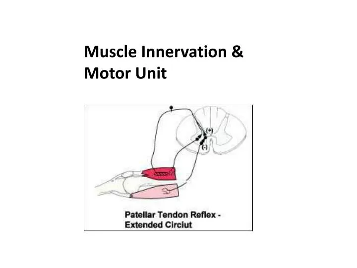
Role of the Muscle Spindle in the Stretch Reflex When the muscle spindle is suddenly stretched by briefly applying a reflex hammer to a tendon, Ia afferents propagate a train of action potentials towards excitatory synapses on alpha motor neurons of homonymous and synergist muscles. These muscles contract immediately and cause a rapid movement. The Ia afferents also excite interneurons that inhibit firing of the alpha motor neurons of the antagonist muscle - a process called reciprocal innervation. The roles of the muscle spindle are: · participating in stretch reflexes, providing proprioceptive information to the CNS about head, trunk and limb position and helping to regulate muscle contraction under the influence of descending motor pathways and afferent inputs.
Reflexes Monosynaptic Reflex also known as Deep Tendon Reflex or Myotactic Reflex · Afferent and efferent limb are directly connected (one synapse) Polysynaptic reflex · Afferent and efferent limb are connected with one or more interneurons Reflex arc components · Sensory receptor (e.g., muscle spindle or Golgi tendon organ) · Afferent neuron · Synapse on efferentneuron (LMN) · Muscle
Stretch Reflex All muscles are usually always under some degree of stretch, so this reflex circuit is normally responsible for the so called ‘muscle tone’. Muscular tone reflects a sustained level of contraction of extrafusal muscular fibers. The stretch reflex is a feedback loop that maintains the muscle length and hence dictates postures. Descending motor pathways (e.g., corticospinal) comprising upper motor neurons can control muscle tone and hence posture by influencing components of the reflex arc.
Landmarks for Reflex Testing · C5, C6: Biceps, brachioradialis · C7, C8: Triceps · L2, L3, L4: Knee jerk · S1: Ankle jerk Scale for Reflex Testing · 4+ Very brisk or hyperactive with clonus. · 3+ Brisker than average (high normal). · 2+ Average (normal). · 1+ Somewhat diminished (low normal). · No response.
. Golgi Tendon Organ These sensory organs are located between tendon and extrafusal muscle fibers. They are in series with extrafusal muscle fibers and are sensitive to the tension developed within the muscle stemming from contraction. Structure of Golgi Tendon Organ Each Golgi tendon organ is innervated by group Ib of muscle afferents Physiological Role of the Golgi Tendon Organ The Golgi tendon organ monitors muscle tension. Golgi tendon organ is part of a reflex that relaxes the homonymous and synergistic muscles and excites the antagonist muscles. This protective reflex is often called the inverse myotactic reflex. The Ib afferent has excitatory synapses on interneurons that in turn inhibit the alpha neurons of the homonymous muscle and other interneurons that excite the alpha motor neurons of the antagonist muscle.
Inverse Stretch Reflex The inverse stretch reflex or inverse myotactic reflex is also called the Golgi tendon reflex is bisynaptic (not monosynaptic), with the second synapse The inverse stretch reflex has two functions: · Protection of the muscle and tendon. Through increased tension within the tendon, the contraction of the muscle is inhibited (via negative feedback). · Maintenance of posture.
Flexion Crossed Extension Reflex • Noxious stimulation of the skin induces a polysynaptic reflex which Produces ipsilateral excitation of Flexor muscles (and inhibition of extensors) and contralateral excitation of extensor muscles (and inhibition of flexors). • Afferent limb: nociceptive fibers (A delta and C). • CNS units: a number of excitatory and inhibitory interneurons over many spinal segments. • Efferent limbs: alpha motor neurons, ipsilateral flexors and contralateral extensors.
Plantar response The normal, adult response is characterized by flexion of the toes (this may be difficult to observe if the flexion crossed extension reflex is vigorously expressed). • Abnormal plantar response: (normal in children up to two years of age) dorsiflexion of the big toe, often accompanied by fanning of the other toes. This type of response is called Babinski’s sign and often indicates a lesion in the corticospinal tract. Hyperreflexia Hyperactive reflexes and clonus suggest an upper motor neuron lesion. Hyporeflexia Possible causes for hyporeflexia are: · Lesion of the spinal segment, Lesion of the LMN, Diseases of the NMJ, Muscle lesions, Sensory loss, Peripheral nerve lesion
Basal Ganglia Location of the Basal Ganglia subcortical islands of gray matter (basal ganglia) in the cerebral white matter. · The most obvious of these structures are the caudate and the putamen ( collectively called the striatum) and the globuspallidus (with the putamen and the globuspallidus collectively forming the lentiform nucleus).
Functional Relations among Structures The basal ganglia modulate motor-related activity of the frontal cortices. Although not directly influencing lower motor neurons, the structures listed below interact to render the basal nuclei as integral to the initiation and control of voluntary movement, owing to their regulation of the thalamus. 1. Striatum: Caudate, putamen 2. Globuspallidus: External (lateral) part (GPe), internal (medial) part GPi) 3. Subthalamic nucleus 4. SubstantiaNigra: Pars reticulata (SNr) and pars compacta (SNc) The principal product of the basal ganglia is an inhibitory signal transmitted to the thalamus, which suppresses an excitatory thalamo-cortical projection. The inhibitory basal gangliar output varies to modulate cortex-dependent motor activity. Because lesions affecting the basal ganglia commonly disturb basal gangliar output and hence cortical activity, such lesions often yield disorders of movement.
Pathways in the basal ganglia Almost all areas of the cerebral cortex project topographically onto the striatum, including a critical input from the motor cortex. The striatum then communicates with the thalamus and then back to the cortex via two different pathways.
Indirect pathway. the striatum has inhibitory input to the external segment of the globuspallidus, which has inhibitory input to the subthalamic nuclei. The subthalamic nuclei project excitatory input to the internal segment of the globuspallidus and the pars reticulata of the substantianigra, which send inhibitory input to the thalamus. The thalamus then sends excitatory input back to the motor cortex. In this pathway, the inhibitory neurotransmitter is GABA, and the excitatory neurotransmitter is glutamate. The overall output of the indirect pathway is inhibitory.
Direct pathway. the striatum sends inhibitory input to the internal segment of the globuspallidus and the pars reticulata of the substantianigra, which send inhibitory input to the thalamus. As in the indirect pathway, the thalamus sends excitatory input back to the motor cortex. Again, the inhibitory neurotransmitter is GABA, and the excitatory neurotransmitter is glutamate. The overall output of the direct pathway is excitatory.