Electromyogram (EMG) Defined
Electromyogram (EMG) Defined. Electromyography (EMG) is an electrical recording of muscle activity which aids in the diagnosis of neuromuscular disease Electrodes Needle Surface. Electromygram (EMG) Procedure. Fine needle is inserted into the muscle to be tested.
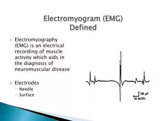

Electromyogram (EMG) Defined
E N D
Presentation Transcript
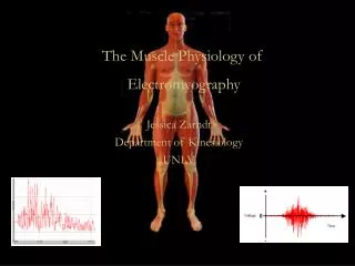
Electromyogram (EMG)Defined • Electromyography (EMG) is an electrical recording of muscle activity which aids in the diagnosis of neuromuscular disease • Electrodes • Needle • Surface
Electromygram (EMG)Procedure • Fine needle is inserted into the muscle to be tested. • Each muscle fiber that contracts will produce an action potential • Presence, size, and shape of the wave form of the action potential are recorded • Recordings are made while the muscle is at rest, and then during the contraction
Electromyography (EMG)Parameters Recorded • Amplitude = negative peak to positive peak • Duration = time from first deflection of the baseline to the last return to baseline • Number of phases = number of times the components of the motor unit potential cross the baseline plus one • Rise time = elapsed time between the peak of the initial positive (down) deflection to the peak of the highest negative (up) deflection Note: the number of fibers contained in a motor unit and their degree of synchrony affect these characteristics
Electromyography (EMG)Insertional Activity • Insertional activity = response of the muscle fibers to needle electrode insertion • Normally consists of brief, transient muscle action potentials in the form of spikes, lasting only a few seconds and stopping immediately when needle movements stop • Abnormal insertional activity: • Decreased • Fibrosis • Fat tissue replacement • Increased • Early denervation • Myotonic disorders
Electromyography (EMG)Spontaneous Activity • Persistence of any activity beyond insertion constitutes spontaneous activity • Could be due to the normal end-plate noise, or to the presence of fibrillations and positive waves, or other spontaneous activity • Normally, the monophasic potentials are of low amplitude and short duration and cause a "thickened baseline" appearance. They give a typical "sea shell" noise or "roar" on the loudspeaker.
Electromyography (EMG)Pathology in Denervated Muscle Fibrillations and Positive Sharp Waves occur with denervation because: • The acetylcholine receptors spread all across the muscle fiber instead of being grouped in the end-plate region • This spread may play a role in attracting new innervation to the denervated muscle fiber from adjacent nerve sprouts • The muscle fiber becomes much more sensitive to free acetylcholine released spontaneously from adjacent nerve fibers and is depolarized and repolarized spontaneously as these molecules reach it • Each single depolarization is electrically detected as a single muscle fiber action potential.
Electromyography (EMG)Abnormal – Fribrillation Potential • Of short duration (<3 msec) and low amplitude (<300 µv), fibrillation potentials occur in semirhythmical runs (<30/second), though occasionally the frequency is so slow it appears to be random. • Develop two to three weeks after the neuron or axon has been damaged • Less frequently seen as time goes by and may be seen infrequently after three years. • As the muscle is reinnervated, both fibrillations and positive waves decrease in number and eventually disappear • Cannot be detected visually on the skin
Electromyography (EMG)Abnormal – Positive Sharp Wave • Very sharp positive deflection off the baseline followed by a slower return and often a negative phase before returning to the baseline • May reach up to 1 mv in amplitude and can last up to 50 msec • Discharge in a very rhythmic manner • Usually the rhythm starts and stops abruptly, and rarely does the individual rhythm vary
Electromyography (EMG)Abnormal - Fasciculation • Spontaneous discharge of an entire unit in a random fashion • Like a cramp • Looks like any motor unit, but is distinguished by the irregular discharge pattern • Can be detected visually on the skin • Binine: regular, normal response
Electromyography (EMG)Repetitive Discharges • A.K.A. high frequency discharges and bizarre repetitive potentials • long trains of rapidly firing potentials with abrupt onset and termination • Seen in a variety of myopathic and neuropathic conditions. • Polymyositis(Polio) • early active stages of Duchenne muscular dystrophy • chronic root lesions • peripheral neuropathies • motor neuron diseases • nerve regeneration
Electromyography (EMG)Myopathic Lesions • Result: unstable spread of the depolarizing current, causing considerable desynchronization in the motor units. • Typically these motor units are of low amplitude, short duration, and have a high number of phases. • In most myopathic lesions neurons remain intact while muscle fibers die or become diseased • This results in: • reduced duration of the motor unit activation • drop in its amplitude • Remaining muscle fibers will do one of the following: • Atrophy • Divide • Separate into small fragments • Split along their axes
Electromyogram (EMG)Normal Response / Values • Muscle tissue is normally electrically silent at rest. • Once the insertion activity quiets down, there should be no action potential on the oscilloscope. • As voluntary contraction is increased, more and more muscle fibers produce action potentials until a disorderly group of action potentials of varying rates and amplitudes (complete recruitment and interference pattern) appears with full contraction. • Voluntary contraction will generate a characteristic biphasic response, i.e. a positive phase followed by a negative one • The rise time, strictly a function of the proximity of the needle tip to the muscle fibers of the contracting unit, is usually between 200 and 300 µsec.
Electromyography (EMG)Primary Uses • Muscular dystrophy • Congenital myopathies • Mitochondrial myopathies-energy making parts • Metabolic myopathies • Myotonias • Peripheral neuropathies • Radiculopathies • Nerve lesions • Amyotrophic lateral sclerosis=Luegarics disease • Polio • Spinal muscular atrophy • Guillain-Barré syndrome • Ataxias • Myasthenias
Nerve Conduction Velocity Studies (NCVs) • Performed to evaluate nerve function and localize site of involvement • Tests the velocity at which impulses travel through a nerve • Two types of NCVs • Motor: stimulate nerve and record over muscle belly • Proximal to distal • Sensory: stimulate sensory nerve and record sensory nerve (not common motor-sensory nerve) • Distal to Proximal • Most are recorded orthodromically (in normal signal direction), though some are recorded antidromically (opposite normal signal direction)
Nerve Conduction Velocity Studies (NCVs) • Nerve is stimulated, usually with surface electrodes. One electrode stimulates the nerve with a very mild electrical impulse. • Resulting electrical activity is recorded by the other electrodes. • Distance between electrodes and the time it takes for electrical impulses to travel between electrodes are used to calculate the nerve conduction velocity.
Nerve Conduction Velocity Studies (NCVs)Procedure • Evoked potentials may also be performed for additional diagnostic information. • NCVs are especially helpful when pain or sensory complaints are more prominent than weakness • Impulse given may feel like a mild electric shock. • Pt. says it hurrts • To stimulate nerves deep to the skin you must use an insulated needle electrode with its uninsulated tip lodged near the nerve.
Nerve Conduction Velocity Studies (NCVs)Motor Procedure • Procedure • Supramaximal impulse is applied eliciting full contraction of muscles distal to stimulus • Typically measured at two different locations and calculated together using equation • M-wave = summated activity of all motor units in the muscle recorded • Latency = time between stimulus and onset of M-wave
Nerve Conduction Velocity Studies (NCVs)Motor Response M-wave represents the summated activity of all motor units (some motor units will be recruited later than others due to slower conduction times), therefore amplitude and shape of wave are important M-wave onset Stimulus Baseline
Nerve Conduction Velocity Studies (NCVs)Normal Values: General Comments • NCV depends on: • Diameter of nerve • Larger =Faster (Sensory) • Degree of myelination • Newborn infants have values that are approximately one-half that of adults, and adult values are normally reached by age 5 • Because haven’t finished myelination yet, periphery at age 5, CNS in teens • Significant decreases in NCVs after age 70 • Demyelination • Specific values available in tables
Nerve Conduction Velocity Studies (NCVs)Normal Values Motor Values • UE values • Average is 60 m/s • Range is 45-70 m/s • LE values • Average is 50 m/s Sensory Values • Typically between 45-75 m/s • Usually sharp wave, unlike rounded M-wave • Slightly faster than motor NCVs because of large diameter sensory nerves
Nerve Conduction Velocity Studies (NCVs)Abnormal Values • Abnormal results may be from: • Demyelination (destruction of the myelin sheath) • Conduction block (the impulse is blocked somewhere along the nerve pathway) • Axonopathy (damage to the nerve axon) • Why we do test in 2 different places, to detect a more distal or proximal lesion.
Nerve Conduction Velocity Studies (NCVs)Primary Uses • Alcoholic neuropathy • Diabetic neuropathy • Nerve effects of uremia (from kidney failure) • Traumatic injury to a nerve • Guillain-Barre syndrome • Diphtheria • Carpal tunnel syndrome • Brachial plexopathy • Charcot-Marie-Tooth disease (hereditary) • Chronic inflammatory polyneuropathy • Common peroneal nerve dysfunction • Distal median nerve dysfunction • Femoral nerve dysfunction
H-ReflexDefined • = Hoffmann Reflex • The H Reflex results from stimulation of 1A afferent fibers with the resulting afferent discharge causing an excitatory potential in the motor neuron pool and muscle activation • Latency of response is a measure of integrity of both sensory and motor fibers
H – ReflexProcedure • Submaximal stimulus applied to S1 nerve roots at tibial nerve in popliteal fossa • Not pictured here • Motor response recorded in medial soleus • Sometimes done in C6-C7 • Pictured here
H – ReflexResponses • NORMAL average response is 29.8ms (+ 2.74 ms) • ABNORMAL responses • Slowed latency abnormal dorsal root function from herniated disk or impingement syndrome • Peripheral motor and sensory NCVs are typically normal in this situation • This test shows abnormalities before EMG denervation potentials would be present
H – ReflexPrimary Uses • Radiculopathy • Peripheral neuropathy
F WaveDefined • A measure of motor neuron conduction • Supramaximal stimulus of motor neurons at a distal site leading to both orthodromic (get distal muscle contraction) and antidromic impulses (goes to anterior horn cell reverberates there impulse sent back down motor neuron recorded) • Antidromic portion of response is response that is called the F wave
F WaveNormal Values (Latencies) • Upper Extremity • Approximately 30 seconds • Lower Extremity • Less than 60 seconds
F WavePrimary Uses • Conditions where proximal nerve is involved • Guillain-Barre Syndrome • Thoracic Outlet Syndrome: UE • Brachial Plexus injuries • Radiculopathies with more than one nerve root involved • As measure of alpha motor neuron excitability in research studies
UltrasoundDefined • Propagated sound waves interact with tissue interfaces to produce images based on reflection or refraction of structures with different acoustic impedance • For Deep Vein Thrombosis • Sound waves are reflected back to a transducer crystal and converted into electrical input • Doppler ultrasound technique produces color-coded real-time images of blood flow.
UltrasoundAdvantages / Disadvantages • Advantages • noninvasive • relatively low cost • Safe, with no radiation • Quick • allows localization of lesions in three dimensions, therefore useful for guiding percutaneous aspiration or biopsy and for mapping radiation portals • Disadvantages
UltrasoundPrimary Uses • Superficial tendons and muscles • Popliteal space • Patellar tendon • Many joints • Popliteal cysts • Tumors and infections of bone and soft tissue • Foreign bodies • Parathyroid glands • Hematomas
Echocardiography • Cardiac imaging technique based upon the velocity of sound traveling through and reflected from acoustic interfaces in cardiovascular structures • Most frequently performed diagnostic study for cardiac diseases • 2-D format most typically used • Doppler format used to examine blood flow through the heart • Transthoracic typically performed • Transesophageal echocardiography involves placement of the ultrasound transducer into the esophagus in proximity to the heart and is sometimes done during cardiac surgeries
EchocardiographyAdvantages / Disadvantages • Advantages • Non-invasive (other than the transesophegeal form) • Readily available
EchocardiographyPrimary Uses • Blood flow mapping of the heart and its blood vessels • Transesophageal echocardiography • imaging of the heart during and after cardiac surgery in the operating room • Stress echocardiography involves the evaluation of regional wall motion following a pharmaceutical stress
Arthrography • Contrast opacification of joint cavities which are then recorded by fluoroscopy, CT, or digital radiography • Application of stress is useful in arthrographic evaluation of ligamentous injuries of the ankle, wrist and first metacarpophalangeal joint.
ArthrographyAdvantages / Disadvantages • Advantages • Can apply stress to a joint during imaging • Good soft tissue images • Disadvantages • Need to inject a radio-opaque substance into joint
Hip developmental dysplasia septic arthritis in infants, Legg Calvè Perthes disease traumatic injuries soft tissue masses Knee (rarely done now since advent of MRI) Ankle ArthographyPrimary Uses • Wrist • Elbow • Glenohumeral • rotator cuff tears • adhesive capsulitis • bicipital tendon abnormalities • rheumatoid arthritis • septic arthritis
Bone Scan • Produced using radiopharmaceutical agents • Shows metabolism of bone • Increased uptake of the radionuclide agent at sites of bone abnormalities • Typically imaged with single photon emission computed tomography (SPECT) • May be imaged with PET scan
Disadvantages Not specific since any process involving changes in bone production and resorption can cause abnormalities on bone scans Bone ScanAdvantages / Disadvantages • Advantages • Very sensitive
Bone ScanPrimary Uses • Bone metastases • Osteomyelitis • Ischemic necrosis of bone • Differentiating osteomyelitis from cellulitis
References • Gale Encyclopedia of Medicine • http://www.nlm.nih.gov/medlineplus/ency • Dorland’s Medical Dictionary • http://www.teleemg.com/Chapters/jbr110.htm • http://www.hucmlrc.howard.edu/neuroanat/Lectures/funanatspincrd.htm