Electromyography (EMG) Instrumentation
Electromyography (EMG) Instrumentation. David Groh University of Nevada – Las Vegas. Research Applications of Surface EMG. Indicator for muscle activation/deactivation Relationship of force/EMG signal Use of EMG signal as a fatigue index. Types of EMG. Electrode Categories Inserted

Electromyography (EMG) Instrumentation
E N D
Presentation Transcript
Electromyography (EMG) Instrumentation David Groh University of Nevada – Las Vegas
Research Applications of Surface EMG • Indicator for muscle activation/deactivation • Relationship of force/EMG signal • Use of EMG signal as a fatigue index
Types of EMG • Electrode Categories • Inserted • Fine-wire (Intra-muscular) • Needle • Surface
Fine-wire Electrodes • Advantages • Extremely sensitive • Record single muscle activity • Access to deep musculature • Little cross-talk concern • Disadvantages • Extremely sensitive • Requires medical personnel, certification • Repositioning nearly impossible • Detection area may not be representative of entire muscle
Surface Electrodes • Advantages • Quick, easy to apply • No medical supervision, required certification • Minimal discomfort • Disadvantages • Generally used only for superficial muscles • Cross-talk concerns • No standard electrode placement • May affect movement patterns of subject • Limitations with recording dynamic muscle activity
Electrode Comparison Studies • Giroux & Lamontagne - Electromyogr. Clin. Neurophysiol., 1990 • Purpose: to compare EMG surface electrodes and intramuscular wire electrodes for isometric and dynamic contractions • Results • No significant difference in either isometric or dynamic conditions • However: dynamic activity was not very “dynamic”
EMG Manufacturers • Noraxon • Motion Lab Systems • Delsys
General Concerns • Signal-to-noise ratio • Ratio of energy of EMG signal divided by energy of noise signal • Distortion of the signal • EMG signal should be altered as minimally as possible for accurate representation
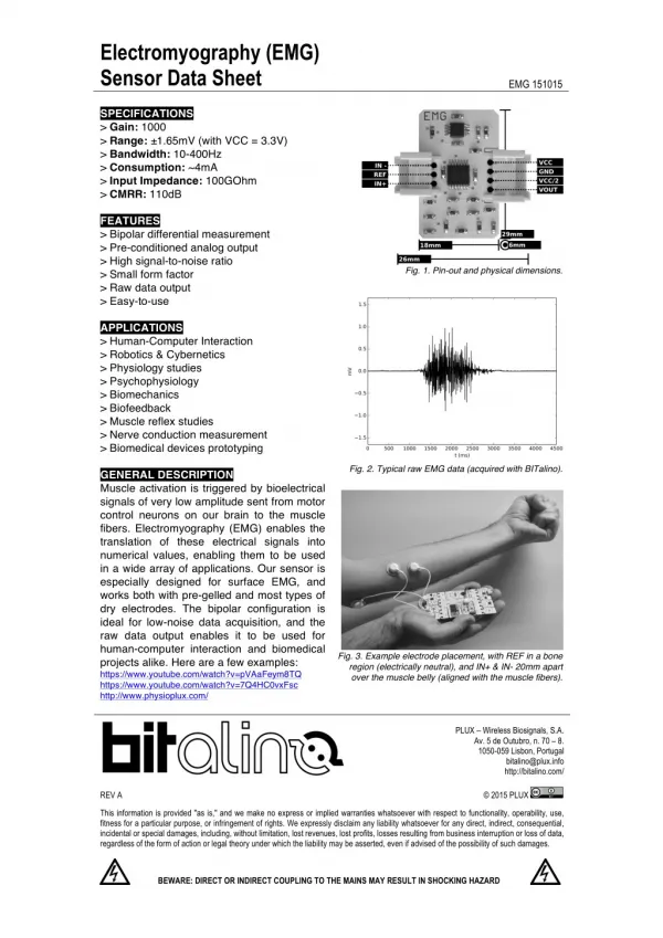
Characteristics of EMG Signal • Amplitude range: 0–10 mV (+5 to -5) prior to amplification • Useable energy: Range of 0 - 500 Hz • Dominant energy: 50 – 150 Hz
Characteristics of Electrical Noise • Inherent noise in electronics equipment • Ambient noise • Motion artifact • Inherent instability of signal
Inherent Noise in Electronics Equipment • Generated by all electronics equipment • Frequency range: 0 – several thousand Hz • Cannot be eliminated • Reduced by using high quality components
Ambient Noise • Electromagnetic radiation sources • Radio transmission • Electrical wires • Fluorescent lights • Essentially impossible to avoid • Dominant frequency: 60 Hz • Amplitude: 1 – 3x EMG signal
Motion Artifact • Two main sources • Electrode/skin interface • Electrode cable • Reducible by proper circuitry and set-up • Frequency range: 0 – 20 Hz
Inherent Instability of Signal • Amplitude is somewhat random in nature • Frequency range of 0 – 20 Hz is especially unstable • Therefore, removal of this range is recommended
Factors Affecting the EMG Signal • Carlo De Luca • Causative Factors – direct affect on signal • Extrinsic – electrode structure and placement • Intrinsic – physiological, anatomical, biochemical • Intermediate Factors – physical & physiological phenomena influenced by one or more causative factors • Deterministic Factors – influenced by intermediate factors
Maximizing Quality of EMG Signal • Signal-to-noise ratio • Highest amount of information from EMG signal as possible • Minimum amount of noise contamination • As minimal distortion of EMG signal as possible • No unnecessary filtering • No distortion of signal peaks • No notch filters recommended • Ex: 60 Hz
Solutions for Signal Interruption Related to Electrode and Amplifier Design • Differential amplification • Reduces electromagnetic radiation noise • Dual electrodes • Electrode stability • Time for chemical reaction to stabilize • Important factors: electrode movement, perspiration, humidity changes • Improved quality of electrodes • Less need for skin abrasion, hair removal
Differential Amplification • Ambient (electromagnetic) noise is constant • System subtracts two signals • Resultant difference is amplified • Double differential technique
Electrode Configuration • Length of electrodes • # of included fibers vs. increased noise*** • Delsys – 1 cm • Noraxon - ? • Distance between electrodes • Increased amplitude vs. misaligning electrodes, Multiple motor unit action potentials (MUAP) • Muscle fibers of motor units are distributed evenly, thus large muscle coverage is not necessary (De Luca). • Delsys – 1 cm • Noraxon – 2 cm?
Electrode Placement • Away from motor point • MUAP traveling in opposite directions • Simultaneous (+) & (-) AP’s • Resultant increased frequency components • More jagged signal • Middle of muscle belly is generally accepted
Electrode Placement • Away from tendon • Fewer, thinner muscle fibers • Closer to other muscle origins, insertions • More susceptible to cross-talk • Away from outer edge of muscle • Closer to other musculature • Orientation parallel to muscle fibers • More accurate conduction velocity • Increased probability of detecting same signal
Reference Electrode Placement(Ground) • As far away as possible from recording electrodes • Electrically neutral tissue • Bony prominence • Good electrical contact • Larger size • Good adhesive properties
References • Basmajian JV, De Luca CJ. Muscles Alive: their functions revealed by electromyography (fifth ed.). Williams & Wilkins, Baltimore, Maryland, 1985 • Cram JR, Kasman GS. Introduction to surface electromyography. Aspen Publishers, Inc. Gaithersburg, Maryland, 1998 • De Luca CJ: Surface electromyography: detection and recording. DelSys, Inc., 2002 • De Luca CJ: The use of surface electromyography in biomechanics. J App Biomech 13: 135-163, 1997 • MyoResearch: software for the EMG professional. Scottsdale, Arizona, Noraxon USA, 1996-1999