Download
1 / 9
140 likes | 442 Views
Understand the types of EMG electrodes (e.g., monopolar, bipolar, concentric core, multi-element) used in electromyography, their materials, procedures, and applications in clinical settings.

E N D
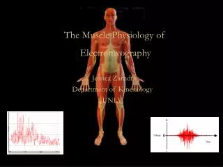
Electromyography (EMG) is a technique for evaluating and recording the electrical activity produced by skeletal muscles. EMG is performed using an instrument called an electromyograph, to produce a record called an electromyogram. • An electromyograph detects the electrical potential generated by muscle cells when these cells are electrically or neurologically activated. The signals can be • analyzed to detect medical abnormalities, activation level, recruitment order or to analyze the biomechanics of human or animal movement.
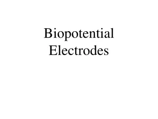
NEEDLE ELECTRODES • Needle electrodes are generally used in clinical electromyography, neurography • and other electrophysiological investigations under the skin and in the deeper • tissues. • Material used: Stainless steel which is preferred due to its mechanical solidity and low price. • These electrodes are generally designed to be fully autoclavable and should be thoroughly sterilized before use • Different types of needle electrodes are used for electromyographic work. They are: Monopolar needle electrodes, Bipolar needle electrodes, Concentric (coaxial) core needle electrode and Multielement needle electrode
MONOPOLAR NEEDLE ELECTRODE • Consists of Teflon coated stainless steel wire which is barely a tip • The coating recedes after being used for several times and the electrode must be discarded when this occurs. They are also color coded.
BIOPOLAR (DOUBLE COAXIAL) NEEDLE ELECTRODE) • Contains two insulated wires within a metal cannula (is a tube that can be inserted into the body, often for the delivery or removal of fluid). • The two wires are bared at the tip and provide the contacts to the patient. • The cannula acts as the ground • These electrodes are electrically symmetrical and have no sense of polarity. The synthesis of a motor unit action potential, as recorded by bipolar needle electrodes
CONCENTRIC(COAXIAL) CORE NEEDLE ELECTRODE • Contains both the active and reference electrode within the same structure. • Consists of an insulated wire contained within an hypodermic needle. • The inner wire is exposed at the tip which forms one electrode. • These needles have very stable electrical characteristics and are convenient to use. • These electrodes are made by moulding fine platinum wire into hypodermic needle having outside diameter less than 0.6mm. • One end is bevelled to expose he end of wire and provide easy penetration. • The surface area of the exposed tip is less than 00005mm sq.
MULTI-ELEMENT NEEDLE ELECTRODES • Used to pick up signals from individual fibers of muscle tissue. • These needles have electrode surfaces of 25 micron diameter and 14 pick up surfaces down the side the side of 1 needle.
GROUND ELECTRODES • Consists of conducting strip which is inserted into a saline soaked strap and wrapped around the patient’s limb. • Usually positioned over bony structures rather than over large muscular masses, in vicinity of the recording and stimulating electrodes, and equidistant from them.