Download
1 / 59
590 likes | 774 Views
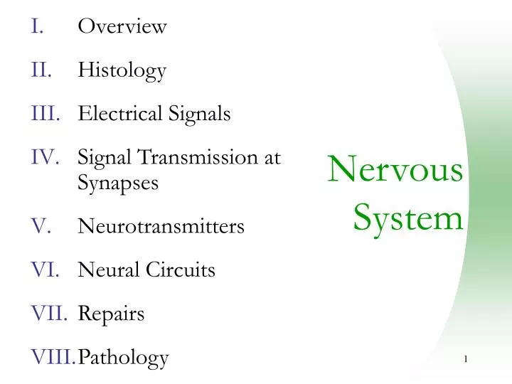
Overview Histology Electrical Signals Signal Transmission at Synapses Neurotransmitters Neural Circuits Repairs Pathology. Nervous System. Overview Structures Functions Organization Histology Electrical Signals Signal Transmission at Synapses Neurotransmitters Neural Circuits

E N D
Overview Histology Electrical Signals Signal Transmission at Synapses Neurotransmitters Neural Circuits Repairs Pathology Nervous System
Overview Structures Functions Organization Histology Electrical Signals Signal Transmission at Synapses Neurotransmitters Neural Circuits Repairs Pathology Nervous System
Nervous Tissue • Controls and integrates all body activities within limits that maintain life • Three basic functions • sensing changes with sensory receptors • interpreting and remembering those changes • reacting to those changes with effectors
Major Structures of the Nervous System • Brain • spinal cord • cranial nerves • spinal nerves • ganglia • enteric plexuses • sensory receptors
Subdivisions of the PNS • Central nervous system (CNS) • Peripheral nervous system (PNS) • Somatic (voluntary) nervous system (SNS) • Autonomic (involuntary) nervous systems (ANS) • Enteric nervous system (ENS)
Organization Sensory Integration Motor SNS (Motor) Brain Spinal cord SNS (Sensory) ANS (Sensory) ANS (Motor) ENS (Sensory)
Overview Histology Neurons Neuroglia CNS PNS Myelination Gray and White Matter Electrical Signals Signal Transmission at Synapses Neurotransmitters Neural Circuits Repairs Pathology Nervous System
Neurons • Functional unit of nervous system • Cell body • Nissl bodies • Neurofilaments • Microtubules • Lipofuscin pigment clumps • Cell processes • Dendrites • Axons
Dendrites impulse • Conducts impulses towards the cell body • Typically short, highly branched & unmyelinated • Surfaces specialized for contact with other neurons • Contains neurofibrils & Nissl bodies
Axons • Conduct impulses away from cell body • Long, thin cylindrical process of cell • Arises at axon hillock • Impulses arise from initial segment (trigger zone) • Side branches (collaterals) end in fine processes called axon terminals • Swollen tips called synaptic end bulbs contain vesicles filled with neurotransmitters
Structural Classification of Neurons • Based on number of processes found on cell body • multipolar = several dendrites & one axon • most common cell type • bipolar neurons = one main dendrite & one axon • found in retina, inner ear & olfactory • unipolar neurons = one process only(develops from a bipolar) • are always sensory neurons
Structural Classification of Neurons • Based on number of processes found on cell body • multipolar = several dendrites & one axon • most common cell type • bipolar neurons = one main dendrite & one axon • found in retina, inner ear & olfactory • unipolar neurons = one process only(develops from a bipolar) • are always sensory neurons
Structural Classification of Neurons • Based on number of processes found on cell body • multipolar = several dendrites & one axon • most common cell type • bipolar neurons = one main dendrite & one axon • found in retina, inner ear & olfactory • unipolar neurons = one process only(develops from a bipolar) • are always sensory neurons
Association or Interneurons • Named for histologist that first described them or their appearance
Neuroglial Cells • Half of the volume of the CNS • Smaller cells than neurons • 50X more numerous • Cells can divide • rapid mitosis in tumor formation (gliomas) • 4 cell types in CNS • astrocytes, oligodendrocytes, microglia & ependymal • 2 cell types in PNS • schwann and satellite cells
Neuroglial Cells (CNS): Astrocytes • Star-shaped cells • Form blood-brain barrier by covering blood capillaries • Metabolize neurotransmitters • Regulate K+ balance • Provide structural support
Neuroglial Cells (CNS): Oligodendrocytes • Most common glial cell type • Each forms myelin sheath around more than one axons in CNS • Analogous to Schwann cells of PNS
Neuroglial Cells (CNS): Microglia • Small cells found near blood vessels • Phagocytic role -- clear away dead cells • Derived from cells that also gave rise to macrophages & monocytes
Neuroglial Cells (CNS): Ependymal cells • Form epithelial membrane lining cerebral cavities & central canal • Produce cerebrospinal fluid (CSF)
Neuroglial Cells (PNS): Satellite Cells • Flat cells surrounding neuronal cell bodies in peripheral ganglia • Support neurons in the PNS ganglia
Neuroglial Cells (PNS): Schwann Cell • Cells encircling PNS axons • Each cell produces part of the myelin sheath surrounding an axon in the PNS
Myelination • Insulation of axon • Increase speed of nerve impulse
Myelination: PNS • All axons surrounded by a lipid & protein covering (myelin sheath) produced by Schwann cells • Neurilemma is cytoplasm & nucleusof Schwann cell • gaps called nodes of Ranvier • Myelinated fibers • Unmyelinated fibers Node of Ranvier
Myelination: PNS • Schwann cells myelinate (wrap around) axons in the PNS during fetal development • Schwann cell cytoplasm & nucleus forms outermost layer of neurolemma with inner portion being the myelin sheath • Tube guides growing axons that are repairing themselves
Myelination: CNS • Oligodendrocytes myelinate axons in the CNS • Broad, flat cell processes wrap about CNS axons, but the cell bodies do not surround the axons • No neurilemma is formed • Little regrowth after injury is possible due to the lack of a distinct tube or neurilemma
Gray and White Matter • White matter = myelinated processes (white in color) • Gray matter = nerve cell bodies, dendrites, axon terminals, bundles of unmyelinated axons and neuroglia (gray color)
Overview Histology Electrical Signals Overview Ion Channels Resting Membrane Potential Graded Potential Generation of Action Potential Propagation of Nerve Impulses Encoding of Stimulus Intensity Comparison of Electrical Signals Signal Transmission at Synapses Neurotransmitters Neural Circuits Repairs Pathology Nervous System
Electrical Signals in Neurons • Neurons are electrically excitable due to the voltage difference across their membrane • Communicate with 2 types of electric signals • action potentials that can travel long distances • graded potentials that are local membrane changes only • In living cells, a flow of ions occurs through ion channels in the cell membrane
Ion Channels • Leakage (nongated) channels are always open • Gated channels canopen and close • Voltage-gated • Chemically (ligand)-gated • Mechanically-gated Na Na
Gated Ion Channels (Voltage-Gated) K K ECF ICF K K K K
Gated Ion Channels (Ligand-Gated) K K NT ECF ICF K K K K
Action Potential Depolarization Na+ • Series of rapidly occurring events that change and then restore the membrane potential of a cell to its resting state • Ion channels open: • Na+ rushes in (depolarization) • K+ rushes out (repolarization) • All-or-none principal • Travels (spreads) over surface of cell without dying out K+ Na+ Repolarization K+
+30 0 -55 -70 Repolarizing Phase of Action Potential Depolarization Na+ • When threshold potential of -55mV is reached, voltage-gated K+ channels open • Na+ channel opening which caused depolarization • K+ channels finally do open, the Na+ channels have already closed (Na+ inflow stops) • K+ outflow returns membrane potential to -70mV causing repolarization • If enough K+ leaves the cell, it will reach a -90mV membrane potential and enter the after-hyperpolarizing phase • K+ channels close and the membrane potential returns to the resting potential of -70mV K+ Hyperpolarization Repolarization
+30 0 -55 -70 Refractory Period of Action Potential Depolarization Na+ • Period of time during which neuron can not generate another action potential • Absolute refractory period • even very strong stimulus willnot begin another AP • Relative refractory period • a suprathreshold stimulus will be able to start an AP K+ Repolarization Hyperpolarization
Propagation of Action Potential • An action potential spreads (propagates) over the surface of the axon membrane • as Na+ flows into the cell during depolarization, the voltage of adjacent areas is effected and their voltage-gated Na+ channels open • self-propagating along the membrane • The traveling action potential is called a nerve impulse A.P. Na Na Na Na Na Na Na Na Na Na
Continuous versus Saltatory Conduction • Continuous conduction (unmyelinated fibers) • Saltatory conduction (myelinated fibers) A.P. Na Na Na Na Na Na Na Na Na Na
Saltatory Conduction • Nerve impulse conduction in which the impulse jumps (Salta) from node to node Na A.P. Na Na Na Na
Overview Histology Electrical Signals Signal Transmission at Synapses Overview Electrical synapses Chemical synapses Excitatoryand Inhibitory Postsynaptic Potential Removal of Neurotransmitter Spatial and Temporal Summation of Postsynaptic Potentials Neurotransmitters Neural Circuits Nervous System
Signal Transmission at Synapses • 2 Types of synapses • electrical • ionic current spreads to next cell through gap junctions • faster, two-way transmission & capable of synchronizing groups of neurons • chemical • one-way information transfer from a presynaptic neuron to a postsynaptic neuron • axodendritic -- from axon to dendrite • axosomatic -- from axon to cell body • axoaxonic -- from axon to axon
Chemical Synapses Presynaptic Neuron • Action potential reaches end bulb and voltage-gated Ca+ 2 channels open • Ca+2 flows inward triggering release of neurotransmitter • Neurotransmitter crosses synaptic cleft & binding to ligand-gated receptors • the more neurotransmitter released the greater the change in potential of the postsynaptic cell • Synaptic delay is 0.5 msec • One-way information transfer Na Na Na Calcium NT Synpatic Cleft NT Postsynaptic Neuron
Excitatory & Inhibitory Potentials Presynaptic Neuron • The effect of a neurotransmitter can be either excitatory or inhibitory • a depolarizing postsynaptic potential is called an EPSP • it results from the opening of ligand-gatedNa+ channels • the postsynaptic cell is more likely to reach threshold • an inhibitory postsynaptic potential is called an IPSP • it results from the opening of ligand-gated Cl- or K+ channels • it causes the postsynaptic cell to become more negative or hyperpolarized • the postsynaptic cell is less likely to reach threshold Na Na Na Calcium NT Synpatic Cleft NT Postsynaptic Neuron Na
Excitatory & Inhibitory Potentials Presynaptic Neuron • The effect of a neurotransmitter can be either excitatory or inhibitory • a depolarizing postsynaptic potential is called an EPSP • it results from the opening of ligand-gated Na+ channels • the postsynaptic cell is more likely to reach threshold • an inhibitory postsynaptic potential is called an IPSP • it results from the opening of ligand-gatedCl- or K+ channels • it causes the postsynaptic cell to become more negative or hyperpolarized • the postsynaptic cell is less likely to reach threshold Na Na Na Calcium NT Synpatic Cleft NT Postsynaptic Neuron K
Removal of Neurotransmitter Presynaptic Neuron • Diffusion • move down concentration gradient • Enzymatic degradation • acetylcholinesterase • Uptake by neurons or glia cells • neurotransmitter transporters Na Na Na Calcium NT Synpatic Cleft NT Postsynaptic Neuron Na K
Summation • Spatial Summation of effects of neurotransmitters released from several end bulbs onto one neuron • Temporal Summation of effect of neurotransmitters released from 2 or more firings of the same end bulb in rapid succession onto a second neuron NT
Summation • Spatial Summation of effects of neurotransmitters released from several end bulbs onto one neuron • Temporal Summation of effect of neurotransmitters released from 2 or more firings of the same end bulb in rapid succession onto a second neuron NT
Summation: Three Possible Responses • Small EPSP occurs • potential reaches -56 mV only • An impulse is generated • threshold was reached • membrane potential of at least -55 mV • IPSP occurs • membrane hyperpolarized • potential drops below -70 mV NT
Overview Histology Electrical Signals Signal Transmission at Synapses Neurotransmitters NT Effect Small-molecule Neuropeptides Neural Circuits Nervous System
Neurotransmitter Effects • Neurotransmitter effects can be modified • synthesis can be stimulated or inhibited • release can be blocked or enhanced • removal can be stimulated or blocked • receptor site can be blocked or activated • Categorized by size: • Small – molecule • Large Synthesis & Release NT Removal Receptor
Small-Molecule Neurotransmitters • Acetylcholine (ACh) • released by many PNS neurons & some CNS • excitatory on NMJ but inhibitory at others • inactivated by acetylcholinesterase Neuron Ach Muscle
Small-Molecule Neurotransmitters • Amino Acids • Glutamate released by nearly all excitatory neurons in the brain • GABA is inhibitory neurotransmitter for 1/3 of all brain synapses • Valium is a GABA agonist - enhancing its inhibitory effect Glutamate (+) GABA (-) Valium