Download
1 / 32
340 likes | 1.13k Views
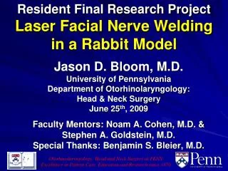
Otorhinolaryngology: Head and Neck Surgery at PENN Excellence in Patient Care, Education and Research since 1870. Resident Final Research Project Laser Facial Nerve Welding in a Rabbit Model. Jason D. Bloom, M.D. University of Pennsylvania Department of Otorhinolaryngology :

E N D
Otorhinolaryngology: Head and Neck Surgery at PENN Excellence in Patient Care, Education and Research since 1870 Resident Final Research Project Laser Facial Nerve Welding in a Rabbit Model Jason D. Bloom, M.D. University of Pennsylvania Department of Otorhinolaryngology: Head & Neck Surgery June 25th, 2009 Faculty Mentors: Noam A. Cohen, M.D. & Stephen A. Goldstein, M.D. Special Thanks: Benjamin S. Bleier, M.D.
Research Approval & Funding • This research project has been approved by both the U of Penn IACUC (Protocol # 802421) & Philadelphia VA ACORP • Awarded 2009-2010AAO-HNSF / AAFPRS Leslie Bernstein Resident Research CORE Grant
Background • Charles Gould (1957) - Introduces the term LASER (Light Amplification by Stimulated Emission of Radiation) • In his doctoral thesis on energy levels of excited thallium • Sigel & Acevado (1962) - Reported thermal energy in tissue adhesion • Passing high frequency electric current through coapted vein edges to anastomose end-to-side porto-caval shunts in dogs (1) • Yahr & Strully (1966) – First description of laser tissue welding in blood vessel anastomosis (2) • Nd-YAG laser • Almquist et al. (1984) – Argon laser in peripheral nerve repair (3) • Fischer et al. (1985) – Published positive reports using the CO2 laser for rat nerve repair (4)
Background Biologic Solder • Allows for target specific laser energy absorption resulting in tissue bonds with greater tensile strength than wounds sealed with laser energy alone (5) • The use of biologic solders has been shown to provide an added benefit in the promotion of native wound healing mechanisms. • Contrast to the granulomatous inflammatory response seen with suture material • The lased solder coagulum provides a non-immunogenic scaffold (6) • Gradually absorbed during the normal wound healing process (7,8) • Biologic solders are now being combined with wavelength specific chromophores (9,10) - Increased target specific energy absorption - Decreased thermal energy leakage - Objective basis of gauging adequacy of laser welding by providing a predictable color change - Examples: Carbon Black 808nm Fluorescein Dye 532nm Indocyanine Green Dye 805nm
Laser Tissue Welding Laser Biologic Solder Chromophore ECM REMODELING LASER WELD
Background • Problems associated w/ classic neurorrhaphy • Trauma induced by handling & sutures • Neuroma formation • Long OR times & technically difficult surgically • “Leakage” of axons at site of anastomosis & in-growth of connective tissue • Foreign body reaction from suture material
Background Laser Nerve Welding • Eppley et al. (1989) – Laser nerve repair of the rabbit facial nerve with CO2 laser (11) • neuroma formation • connective tissue invasion • axonal proliferation or extravasation outside of epineurium • Problems: • Histopathologic weld analysis • Demonstrated some deleterious effects of the heat from the laser, such as destruction of myelin and loss of axons immediately adjacent to the anastomotic site. • Poor anastomosis tensile strength & dehiscence
Background Laser Nerve Welding • Trickett et al. (1997) – Albumin based solder with indocyanine green chromophore to laser weld rat sciatic nerves with an 800nm diode laser (12) - Laser activated solders = stronger bonds at the anastomosis site & less thermal damage to underlying nerve tissue - Histology = denatured solder and underlying epineurium with no axonal damage • Hwang et al. (2005 & 2006) – CO2 laser welding of rat facial nerve w/o solder (13,14) • Laser repaired nerve regeneration = microsuture repaired nerve regeneration immmunohistochemical nerve tracer • Less cellular & fibroblastic response • Less scar & neuroma formation
Hypothesis • Laser welding of the rabbit facial nerve provides a method of creating an atraumatic, immediate and strong repair bond capable of allowing axonal regeneration across the injury site without damage to the nerve itself or the complications associated with suture neurorrhaphy. • Null Hypothesis: Laser nerve welding is not an effective way to perform neurorrhapy, as compared to suture nerve repair.
Rabbit Survival Surgery • 14 New Zealand White Rabbits (3-4kg) • Housed at the Animal Research Facility (ARF) of the Philadelphia Veterans Affairs Medical Center (PVAMC) • 2 of the rabbit facial nerves (negative control group) = facial nerves transected with a 1cm piece of nerve excised w/o re-anastomosis • 6 of the rabbits facial nerves = suture anastomosis w/ three 9-0 monofilament nylon sutures on an atraumatic taper needle • 6 of the rabbit facial nerves = laser welded utilizing an 810nm diode laser and a “ribbon” of 42% albumin-based biologic solder coupled with an indocyanine green dye chromophore applied with a 27 G needle
Identifying the Facial Nerve Hand-held nerve stimulator was usedto identify the facial nerve and verify facial movement. * Also, used to verify that the facial nerve was cut no longer innervating the face
Suture Neurorrhaphy Suture anastomosis w/ three 9-0 monofilament polypropylene (Prolene) sutures on an atraumatic taper needle * Carl Zeiss OPMI CS-1 operating microscope
Laser/Solder Platform Biologic solder - 42% albumin solution Indocyanine green dye Hyaluronic acid sodium Diode laser module - Iridex Oculight SLx Power: 0.5W Pulse Duration: 0.5s Pulse Interval: 0.1s Power Density: 15.9W/cm2 Fluency: 8.0 J/cm2 Major Wavelength Output: 810 +/- 1nm * 600mm core diameter quartz-silica fiberoptic cable
Laser Solder Neurorrhaphy • Microvascular clip – used to hold nerve ends in place • Adequacy of weld was visually assessed by the green colored chromophore changing to a brown color s/p laser energy
Surgical Complications Surgical Complications:Sialocele (5/14 rabbits = 35%) * All treated w/ repeated 20G needle aspirations all completely resolved in 2wks * No pain, distress, feeding issues, effect on wt. gain
EMG Testing Nicolet Viking IV EMG- subdermal electrodes Nerve Conduction Study - measuring CMAPs - amplitude - nerve latency
Functional / Clinical Testing • Positioning of the rabbits that allows standardized visualization and video recording of facial movements. • Gentle nasal & forehead taps performed in an attempt to elicit facial nerve motion. • Video tapes will then be analyzed to objectively grade facial nerve recovery by 2 different blinded observers. • Assess the rabbit's bilateral upper lip & whisker movement. • A scale of facial nerve movement from 0-3 will be used to record this functional testing with 0=no movement and 3=normal facial movement (16). Normal Facial Nerve Function Left Facial Nerve Paralysis
Histological Testing • 6 nerve repairs from each experimental group were harvested and fixed in 10% buffered formalin for subsequent H&E staining • Analysis by our histopathologist (Eugene Einhorn, MD) for immune response to the solder, native wound healing progression, and extent of collateral nerve thermal injury. • The welds were also be prepared and stained with Masson’s Trichrome by our histopathologist, in order to view the axon counts and arrangement of nerve axons across the repair sites. Nerve Laser Weld (H&E 4x) – 4wks * 16 week histological analysis = ongoing (1) Axon counts growing across the nerve repair site (2) Organization of the axons (3) Amount of neuroma formation (4) Amount of foreign body/granulomatous reaction
Operative Time * *P < 0.01 N=6 N=6
Problems with the Study Multiple buccal branches of the rabbit facial nerve distal to the site of the nerve injury & repair Small number of rabbits in each group Inability to circumferentially laser weld the nerve Inconsistent EMG electrode placement in the facial musculature for each separate trial No adequate method to measure synkinesis in a rabbit model Testing EMG vs. ENoG
Discussion / Future Directions • Further areas of research with this laser welding technology: • Peripheral nerve repair • Microvascular arterial or venous anastomosis • Pharyngocutaneous fistula repair • Transoral robotic surgery models • Augmentation of the biologic solder with growth factors and antibiotics to further optimize wound healing • If these welds prove efficacious in larger trials, this technology would be ideally transitioned to clinical trials of facial nerve injury repair.
LNW Conclusions • Clinical / Functional: • Improved facial nerve functional recovery on video analysis with the laser weld group • Electrophysiological: • Improved facial nerve electrophysiological/EMG nerve recovery at all time points in the laser weld group, especially at early (1 month) & late (4 month) time points • Histological: • H&E staining demonstrating negligible collateral thermal nerve or tissue damage • Biologic solder supporting the progression of the native wound healing response • Ongoing histopath: Utilizing Masson’s Trichrome stain (1) Axon counts growing across the nerve repair site (2) Organization of the axons (3) Amount of neuroma formation (4) Amount of foreign body/granulomatous reaction • Mechanical: • No learning curve or operating microscope necessary with the laser weld group • Significantly reduced neurorrhaphy time with the laser weld group
References • 1. Sigel B, Acevado FJ, “Vein anastomosis by electrocoaptive union,” Surg Forum. 1962;13:233-5. • 2. Yahr WZ, Strully KJ, “Blood vessel anastamosis by laser and other biomedical applications,” J Assoc Adv Med Instrum. 1966;(1):28-31. • 3. Almquist EE, Nachemson A, Auth D, Almquist B, Hall S. “Evaluation of the use of the argon laser in repairing rat and primate nerves,” J Hand Surg Am. 1984;9:792-299. • 4. Fischer DW, Beggs JL, Kenshalo DJ, Shetter AG. “Comparative study of microepineurial anastomoses with the use of CO2 laser and suture techniques in rat sciatic nerves: Part 1. Surgical technique, nerve action potentials, and morphological studies,” Neurosurgery. 1985;17:300-308. • 5. Gil Z, Shaham A, Vasilyev T, Brosh T, Forer B, Katzir A, Fliss DM. “Novel laser tissue-soldering technique for dural reconstruction,” J Neurosurg. 2005 Jul;103(1):87-91. • 6. Kirsch AJ, Miller MI, Hensle TW, Chang DT, Shabsigh R, Olsson CA, Connor JP, “Laser tissue soldering in urinary tract reconstruction: first human experience,” Urology. 1995 Aug;46(2):261-6. • 7. Lauto A, Trickett R, Malik R, Dawes JM, Owen ER, “Laser-activated solid protein bands for peripheral nerve repair: an vivo study,” Lasers Surg Med. 1997;21(2):134-41. • 8. Lauto A, Kerman I, Ohebshalon M, Felsen D, Poppas DP, “Two-layer film as a laser soldering biomaterial,” Lasers Surg Med. 1999;25(3):250-6. • 9. Talmor M, Bleustein CB, Poppas DP, “Laser tissue welding: a biotechnological advance for the future,” Arch Facial Plast Surg. 2001 Jul-Sep;3(3):207-13. • 10. Oz MC, Johnson JP, Parangi S, Chuck RS, Marboe CC, Bass LS, Nowygrod R, Treat MR, “Tissue soldering by use of indocyanine green dye-enhanced fibrinogen with the near infrared diode laser,” J Vasc Surg. 1990 May;11(5):718-25. • 11. Eppley BL, Kalenderian E, Winkelmann T, Delfino JJ. “Facial nerve graft repair: suture versus laser-assisted anastomosis,” Int J Oral Maxillofacial Surg. 1989;18:50-54.
References • 12. Trickett I, Dawes JM, Knowles DS, Lanzetta M, Owen ER. “In vitro Laser Nerve Repair: Protein Solder Strip Irradiation or Irradiation Alone?,” Int Surg. 1997;82:38-41. • 13. Hwang K, Kim SG, Kim DJ, Lee CH. “Laser Welding of Rat’s Facial Nerve,” Journal of Craniofacial Surgery. 2005;16(6):1102-1106. • 14. Hwang K, Kim SG, K DJ. “Facial-Hypoglossal Nerve Anastomosis Using Laser Nerve Welding,” Journal of Craniofacial Surgery. 2006;17(4):687-691. • 15. Yian CH, Paniello RC, Spector JG. “Inhibition of Motor Nerve Regeneration in a Rabbit Facial Nerve Model.” Laryngoscope. 2001;111:786-791. • 16. Bleier BS, Palmer JN, Sparano AM, et al. “Laser-assisted cerebrospinal fluid leak repair: an animal model to test feasibility.” Otolaryngol Head Neck Surg. 2007;137(5):810-4. • 17. Bleier BS, Palmer JN, Gratton MA, Cohen NA. “In Vivo Laser Tissue Welding in the Rabbit Maxillary Sinus.” American Journal of Rhinology. 2008;22:625-628. • 18. Bleier BS, Gratton MA, Leibowitz JM, Palmer JN, Newman JG, Cohen NA. “Laser-Welded Endoscopic Endoluminal Repair of Iatrogenic Esophageal Perforation: An Animal Model.” Otolaryngology – Head and Neck Surgery. 2008;139:713-717.
THANK YOU!Special Thanks To:- My Faculty Mentors- Dr. O’Malley- Dr. Cohen, Dr. Palmer, Dr. Bleier & The Penn Rhinology Lab- Perry DeRitis & VA ARF Staff- VA Neurology Service