Download
1 / 25
280 likes | 721 Views
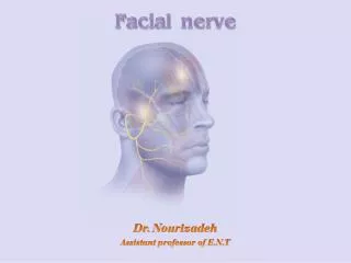
Facial Nerve Disease. Shankai Yin Prof Dept of Otolaryngology, the sixth hospital affiliated to Shanghai jiaotong university Otolaryngology institute at Shanghai jiaotong university. Anatomy. Facial nerve is a mixed nerve, having a motor root and a sensory root.

E N D
Facial Nerve Disease Shankai Yin Prof Dept of Otolaryngology, the sixth hospital affiliated to Shanghai jiaotong university Otolaryngology institute at Shanghai jiaotong university
Anatomy • Facial nerve is a mixed nerve, having a motor root and a sensory root. • Motor root supplies all the mimetic muscles of the face which develop from the 2nd brachial arch.
Sensory root “nerve of Wrisberg” carries taste fibers from the anterior 2/3 of the tongue and general sensation from the concha and retroauricular skin. • Also it carries secretomotor fibers to the lacrimal, submandibular and sublingual glands as well as those in the nose and palate.
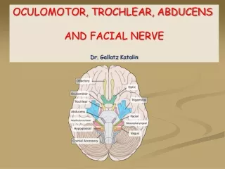
Anatomy: Parts • Intracranial part • Intratemporal part • Extracranial part
Course of the Facial Nerve • Intracranial • Arises at the pontomedullary junction and courses with CNVIII to the internal acoustic meatus • 12mm
Intratemporal • Meatal • Anterior to the superior vestibular nerve and superior to the cochlear nerve – 10mm • Labyrinthe segment • Passes through narrowest part of fallopian canal - 12mm • Narrowest part of facial nerve. The most susceptible to compression secondary to edema. • Tympanic segment • From geniculate ganglion to pyramidal turn – 11mm • Mastoid segment • Exits the stylomastoid foramen – 13mm
Extracranial • From stylomastoid foramen to pesanserinus
Anatomy: Branches • Greater superficial petrosal nerve • Nerve to stapedius • Chorda tympani • Comunicating branch • Posterior auricular nerve • Muscular branches • Peripheral branches: “Pesanserinus”
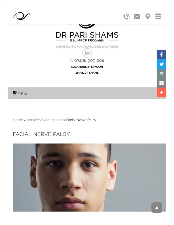
Presentation • Functional and cosmetic problems • Upper lid fails to drop down and close • Lower lid loses tone and sags downward • May evert leading to ectropion • Produces lagophthalmos and consequent corneal exposure. • Interruption of the tear film • Leads to drying of cornea • Ocular discomfort • Corneal ulcers • Infection • Perforation
Upper motor neurone (UMN) • can wrinkle their forehead (unless bilateral lesion) • sagging of the face seen with lower motor neurone palsies is not as prominent. • Lower motor neurone (LMN) • can't wrinkle their forehead
I Normal II Normal tone and symmetry at rest. Slight weakness on close inspection Good to moderate movement of forehead. Complete eye closure with minimum effort. Slight asymmetry of mouth with movement III Normal tone and symmetry at rest. Obvious but not disfiguring facial asymmetry. Synkinesis may be noticeable but not severe .+/- hemifacial spasm or contracture. Slight to moderate movement of forehead Complete eye closure with effort. Slight weakness of mouth with maximum effort. IV Normal tone and symmetry at rest. Asymmetry is disfiguring or results in obvious facial weakness. No perceptible forehead movement. Incomplete eye closure. Asymmetrical motion of mouth with maximum effort V Asymmetrical facial appearance at rest. Slight, barely noticeable movement. No forehead movement. Incomplete eye closure. Asymmetrical motion of mouth with maximum effort. House-Brackmann Facial NerveGrading Scale
Degree of Lesion • Sunderland classification • 1° Partial block: Neuropraxia • 2°Loss of axons: axonotemesis • 3°Injury to the endoneurium: neurotemesis • 4°Injury to the perineurium: partial transection • 5°Injury to the epineurium: complete transection
Diagnosis • History • Presentation • Hearing test • Vestibular function • MRI / CT • Topognostic - Where is the lesion? • Qualitative -Degree of the lesion
Topodiagnostic Diagnosis • Schirmer’s tear test • Stapedius reflex • Taste test • Submandibular salivary flow test
QualitativeDiagnosis • Nerve Excitability Test: NET • Maximum stimulation Test: MST • Electroneurography: ENoG • Electromyography: EMG
Bell’s Palsy • 60-70% cases • Pathophysiology – Impaired “axoplasmic” flow from edema of facial nerve within fallopian canal • Rapid onset and evolution < 48 hours • May be associated with acute neuropathies of cranial nerves V- X • Pain or numbness affecting ear, mid-face, tongue and taste disturbances • Recurrences are more likely (2.5x) in patients with family history, immunodeficiency or diabetes
Pathophysiology • Main cause of Bell's palsy is latent herpes viruses (herpes simplex virus type 1 and herpes zoster virus), which are reactivated from cranial nerve ganglia. • Polymerase chain reaction techniques have isolated herpes virus DNA from the facial nerve during acute palsy.
Treatment • Oral antivirals - Acyclovir - 10mg/kg (500mg) q8hrs x 7 days • Corticosteroid taper 1mg / kg / day for 10 days • Eye protection - lacrilube • Follow progression with serial exams • Facial nerve decompression • Progression to > 90% degeneration on ENOG • Performed before irreversible injury to the endoneurial tubules occurs (two weeks), will allow for axonal regeneration to occur
Herpes Zoster Oticus(Ramsay Hunt syndrome) • 10-15% of acute facial palsy cases • Lesions may involve the external ear, the skin of EAC or soft palate • Associated symptoms – hearing loss, dysacusis and vertigo • Additional involvement of CN V, IX and X and cervical branches 2, 3 and 4 • Pathogenesis – Neural injury due to edema at point between the meatal foramen and the geniculate fossa in the labyrinthe segment