Download
1 / 45
450 likes | 593 Views
Review of Questions on Conformational Changes in Membrane Receptor Rhodopsin and Overview of Bulk Biophysical Methods . Judith Klein-Seetharaman Co-Course Director jks33@pitt.edu. From Last Lecture: Tips on Lecture Preparation. Give a roadmap

E N D
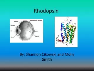
Review of Questions on Conformational Changes in Membrane Receptor RhodopsinandOverview of Bulk Biophysical Methods Judith Klein-SeetharamanCo-Course Director jks33@pitt.edu
From Last Lecture: Tips on Lecture Preparation • Give a roadmap • Give an overview of the field introducing your topic • Use visual aids as much as possible - but is okay to have text if needed • Basic architecture of a slide: • Title • Subtitle • Text and/or image • Conclusion line • Give appropriate references (papers, websites) for resources you used (especially images) • Have a summary at the end of the lecture • Use my last lecture as a template for font sizes and colors (pls. do not use your own style) Molecular Biophysics 3: Lecture 4
From last lecture: Summary Conformational changes in rhodopsin • Introduction to rhodopsin • Seven transmembrane helical bundle with posttranslational modifications and ligand retinal bound • General approach: cysteine mutagenesis and attachment of biophysical probes • Biophysical methods applied to single cysteines • Mobility with EPR spectroscopy • Secondary structure from EPR mobility • Helix orientation from EPR mobility • Tertiary structure from EPR hyperfine lines • Tertiary structure from cysteine reactivity rates with absorbance spectroscopy • Aqueous / membrane boundary from EPR collision frequency (O2 versus NiEDDA) • Exposed loops from cysteine reactivity • Biophysical methods applied to double cysteines • Proximity from disulfide bond formation rates • Proximity from EPR spin spin interactions • Conformational changes by comparing the above parameters for different states – here dark versus light Molecular Biophysics 3: Lecture 4
From last Lecture: Summary Current Picture of Conformational Changes upon Light Activation Picture from proximity studies IV II III Picture from single cysteine studies V I Rigid Body Movement (Biophys. Evidence) vs. Changes in Mobility (Crystallography) VI VII Picture from crystal structure Molecular Biophysics 3: Lecture 4
How would you study the significance of the conformational changes for function? Molecular Biophysics 3: Lecture 4
How would you study the significance of the conformational changes for function?Prevent conformational changes and see if the protein still functions correctly (here binds proteins of the signal transduction cascade) Molecular Biophysics 3: Lecture 4
From Structure/Dynamics to Function Effect of restricting conformational changes on function Phosphor- ylation G T Activation by RK No Effect No Effect Abolished Abolished Enhanced Abolished Conformational changes need not be cooperative, but can be segmental Molecular Biophysics 3: Lecture 4
What other models of activation can you imagine? On blackboard Molecular Biophysics 3: Lecture 4
Models for Signal Transduction Mechanisms Conformational changes “frozen” dynamic signal transduction model mechanical signal transduction models Kim S.-H., Prot.Sci., 3 (1994), pp. 159-165 Ottemann K.M., Science, 285 (1999), pp. 1751-1754 Molecular Biophysics 3: Lecture 4
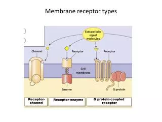
Membrane Receptor Families GPCR Chemo-/Phototaxis EGFR Integrins Two types of receptors: Type I and Type II Molecular Biophysics 3: Lecture 4
Objectives of this Lecture • Overview of bulk methods and what they are used for • Use the topic conformational changes in membrane receptors to gather a list of biophysical methods (your homework) • Decide on who will write summaries on what methods • Limitations of biophysical bulk methods • Go through your questions on conformational changes in rhodopsin – will only have time for one Molecular Biophysics 3: Lecture 4
Overview of methods On blackboard Molecular Biophysics 3: Lecture 4
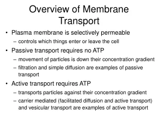
Organized by purpose • To study biomolecular interactions • To study dynamics • To identify secondary structures • To identify oligomerization state • To identify tertiary structures Molecular Biophysics 3: Lecture 4
Overview of bulk methods to study biomolecular interactions • Identification (mass spectrometry, HPLC, absorbance, fluorescence, radioactivity) • Kinetics (absorbance, fluorescence, Biacore) • Size (gel filtration, light-scattering…) • Identification of specific interaction sites (fluorescence quench, spin spin interactions…) • Accessibility • NMR spectroscopy • X-ray crystallography • Cryo-EM Molecular Biophysics 3: Lecture 4
Overview of bulk methods to study dynamics • Global methods: • Methods to identify secondary structure (changes) • Methods to identify tertiary structure (changes) • Methods to quantify oligomerization state • Semi-quantitative methods: • Accessibility at specific sites • Motion at specific sites • Identification of specific tertiary contacts • Atomic resolution methods: • Accessibility by H/D exchange + NMR or mass spec • NMR spectroscopy (structure and dynamics) • X-ray crystallography (mostly structure) • Cryo-electron microscopy (mostly structure) Molecular Biophysics 3: Lecture 4
Techniques to identify secondary structure • CD spectroscopy • FTIR • X-ray crystallography • NMR spectroscopy • EPR spectroscopy (spin label) Molecular Biophysics 3: Lecture 4
Techniques to identify oligomerization state • Gel filtration • Static light scattering • Dynamic light scattering • SANS/SAXS Elution volume [ml] Molecular Biophysics 3: Lecture 4
Techniques to identify tertiary structure • Intrinsic Fluorescence • Fluorescence (fluorescence label) • EPR spectroscopy • FTIR • Raman • CD spectroscopy • (far-UV – secondary structure, near-UV – immobilization of aromatic residues in tertiary structure) • Dye binding (e.g. ANS) • NMR spectroscopy (1H,13C,15N,2H,19F) • Solution • Solid state • X-ray • Cryo-EM Molecular Biophysics 3: Lecture 4
Comparison of qualitative tertiary structure descriptors – complementarity of methods • Motion and tertiary structure indicators • EPR single labels • Cysteine reactivity • Fluorescence labels • NMR labels • Accessibility • EPR collision • 19F NMR • 1H/2H titrations • Cysteine reactivity • Identification of specific tertiary interaction sites • EPR spin spin interaction • NMR (solution or solid-state) spin spin interaction • FRET • Disulfide or chemical crosslinking Molecular Biophysics 3: Lecture 4
Limitations On blackboard Molecular Biophysics 3: Lecture 4
Limitations • Stability • Resolution • Need for labeling • Information content • Native environment • Applicability • Quantities and source of biomolecules Gather a list of limitations on the blackboard Molecular Biophysics 3: Lecture 4
Stability Crystallography of the dark-light transition in rhodopsin Crystals dark Crystals light The conformational changes upon illumination destroy the crystals and crystals grown in the light do not diffract. Molecular Biophysics 3: Lecture 4
2. Resolution In NMR: resolve signals from individual atoms folded unfolded Unfolded proteins have a smaller chemical shift dispersion than folded proteins. Molecular Biophysics 3: Lecture 4
2. Resolution In crystallography: resolve atom positions • Example rhodopsin: In rhodopsin: conformational changes may be smaller than resolution of the crystal Molecular Biophysics 3: Lecture 4
2. Resolution In crystallography: resolve atom positions • Example rhodopsin: conformational changes smaller than resolution of the crystal structure • Example low barrier hydrogen bonds: Certain questions require very high resolution. Molecular Biophysics 3: Lecture 4
2. Resolution Time resolution Time scales of biological processes vary from fs to days. Molecular Biophysics 3: Lecture 4
3. Need for labeling Signal versus background • Intrinsic Fluorescence • no tryptophans, no fluorescence • NMR • No 15N, 13C, no selective excitation • FTIR, Raman • Large background signals • Resonance Raman • Needs a chromophore • Attachment of probes (EPR, fluorescence, etc.) • Perturbation of the native environment by large size Molecular Biophysics 3: Lecture 4
4. Information content • Semi-quantitative methods • Limited information only • X-ray crystallography • Snap shots only • Cryo-EM • Lower resolution Molecular Biophysics 3: Lecture 4
5. Native environment • In vitro studies • Lacks molecular crowding of the cell • Membrane proteins • Lipid bilayer • X-ray crystallography • Not in solution Molecular Biophysics 3: Lecture 4
6. Applicability • X-ray • Crystallization conditions empirical • NMR • Limited by size • Limited by labeling ability • Transient intermediates • adequate time-resolution • Sensitivity to conformational changes • small changes need to be detectable (size of label often much larger than distance change) Molecular Biophysics 3: Lecture 4
Applicability: X-ray Crystallography of the dark-light transition in rhodopsin Crystals dark Crystals light Applicability is also tied in with resolution Molecular Biophysics 3: Lecture 4
Applicability: X-ray So why are we now getting crystals? 2I36 2I37 Molecular Biophysics 3: Lecture 4
Applicability: X-ray So why are we now getting crystals? Alternative explanation: crystal packing artifacts stabilize conformations that are not stable in solution. Molecular Biophysics 3: Lecture 4
Applicability: X-ray Crystallography of the dark-light transition in rhodopsin Crystals dark Crystals light Applicability is also tied in with resolution Molecular Biophysics 3: Lecture 4
So, let’s just do NMR with rhodopsin! Molecular Biophysics 3: Lecture 4
When would you do NMR with a protein? Molecular Biophysics 3: Lecture 4
NMR spectroscopy • Size • Stability • Sample homogeneity • Need for labeling • Quantities and source of biomolecules General limitations Molecular Biophysics 3: Lecture 4
Example Solution NMR of DAGK 1H,15N-HSQC spectrum of a 120 aa long membrane protein in DPC micelles Diacylglycerol kinase: Charles R. Sanders, Frank Sonnichsen (2006) Solution NMR of membrane proteins: practice and challenges. Magn. Reson. Chem. 2006; 44: S24–S40 Molecular Biophysics 3: Lecture 4
Example Solution NMR of Rhodopsin It’s a headache. Molecular Biophysics 3: Lecture 4
What is signal 1? How can you test your hypothesis? Molecular Biophysics 3: Lecture 4
Assignment of Signal 1 NMR Spectroscopy Black: original spectrum, red: C-terminus, green: N-terminus (after AspN cleavage) An enzyme was used to cleave off the C-terminus at the site indicated below: Molecular Biophysics 3: Lecture 4
Traditional Solution NMR Approaches Problems with full-length membrane proteins in detergents • Size – is not the only problem (Trosy does not work for helical membrane proteins) • Conformational exchange – fluctuations in the detergent micelle environment lead to fast relaxation thus signal decay • Spin diffusion – cannot deuterate samples from mammalian cells Problem: Traditional assignment strategies using triple resonance experiments (13C,15N,1H) don’t work Klein-Seetharaman et al. (2004) PNAS 101, 3409-13. Molecular Biophysics 3: Lecture 4
Your questions • If you find time, pls. go through the document that has all the questions and write an answer Conformational Changes in Rhodopsin Molecular Biophysics 3: Lecture 4
Summary of this Lecture Complementarity of biophysical bulk methods Example Membrane Receptor Conformational Changes • Stability • Resolution • Need for labeling • Information content • Native environment • Applicability • Quantities and source of biomolecules Molecular Biophysics 3: Lecture 4
A request Please send word files to Oznur if at all possible, especially with the questions. Molecular Biophysics 3: Lecture 4