Download
1 / 81

E N D
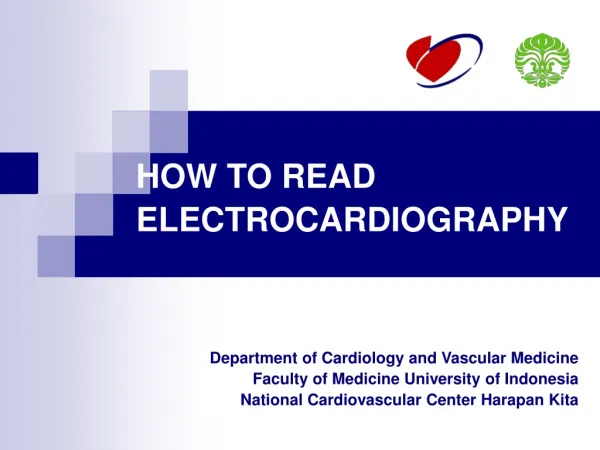
HOW TO READ ELECTROCARDIOGRAPHY Department of Cardiology and Vascular Medicine Faculty of Medicine University of Indonesia National Cardiovascular Center Harapan Kita
Midclavicular line Anterior axillary line Midaxillary line V6 V6R V5 V5R V4 V4R V3 V3R V2 V1 Unipolar Precodial (Chest) Leads Mervin J. Goldman, MD. 11th edition Principles of clinical Electrocardiography. Clinical Professor of Medicine University of California School of Medicine San Francisco @1995-1982
Horizontal plane of V4-6 V7 V8 V9 V9RV8RV7R Unipolar Precodial (Chest) Leads Mervin J. Goldman, MD. 11th edition Principles of clinical Electrocardiography. Clinical Professor of Medicine University of California School of Medicine San Francisco @1995-1982
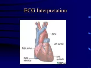
The electrocardiogram (ECG) illustrates conduction of electrical impulses in the heart. The depolarization of the ventricles occurs from the endocardium (inside) to the epicardium (outside) [e] The repolarization of the ventricles occurs in the opposite direction. [g]
ECG INTERPRETATION • RATE • RHYTHM • AXIS • HIPERTROPHIC SIGNS • MYOCARDIAL INFARCTION • ARRHYTHMIA
1. RATE • Normal heart rate : 60 – 100 x/minutes • > 100 x/minutes : Sinus Tachycardia • < 60 x/minutes : Sinus Bradicardia • Determination heart rate (normal paper speed 25 mm/s): • 300 • Count number of large square (bold boxes in one R – R’ interval) • 1500 • Count number of small square in one R – R’ intervals • Number of QRS complex in 6 seconds, multiply by 10
2. RHYTHM Normal cardiac rhythm : SINUS rhythm • Sinus rhythm characteristics : • Rate 60-100 bpm • Constant R – R interval • Negative P wave in aVR and positive di II • P wave is always followed by QRS complex
5. MYOCARDIAL INFARCTION • Ischemia • Injury • Necrosis
WHAT’S WRONG?? Lead Error: V1 and V3 are Transposed! In this normal 12-lead ECG the V1 and V3 chest electrodes are interchanged. Experienced ECG interpreters should be able to spot this lead placement error.