Download
1 / 5
50 likes | 77 Views
Direct Plagiarism

E N D
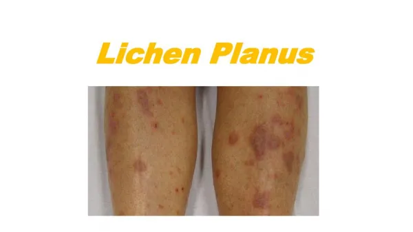
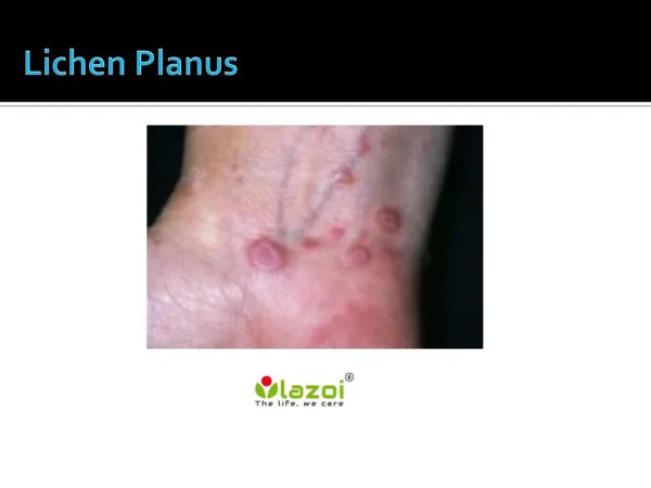
Vol. 79 No. 6 June 1995 Margot L. Van Dis and Edwin T. Parks Article : Prevalence of oral lichen planus in patients with diabetes mellitus ORAL MEDICINE Editor: Jed Jacobson Prevalence of oral lichen planus in patients with diabetes mellitus Margot L. Van Dis, DDS, MS, a and Edwin T. Parks, DMD, MS, b Indianapolis, Ind. DEPARTMENT OF STOMATOLOGY, INDIANA UNIVERSITY SCHOOL OF DENTISTRY The purpose of this study was to determine the prevalence of oral lichen planus in a population of patients with diabetes mellitus compared with a control population and to determine if patient medications had any influence on the presence of such lesions. Two hundred seventy-three patients with diabetes and an identical number of age-, gender- and race-matched controls were examined for clinical evidence of oral lichen planus. Patient medication histories were also obtained from each group. Eleven diabetic patients (4%) and eight control patients (3%) had clinical evidence of oral lichen planus. Ingestion of nonsteroidal anti-inflammatory drugs or angiotensin-converting enzyme inhibitors was associated with the presence of oral lichen planus lesions in six patients. There was no apparent association of diabetes and oral lichen planus in this population, and the ingestion of medications known to cause tichenoid mucosal reactions had no influence per se on the presence of oral lichen planus lesions (p > 0.05). However, the type of medication ingested by those patients who had oral lichen planus lesions was either nonsteroidal anti-inflammatory drug or angiotensin-converting enzyme inhibitor, which was a significant association (t9 < 0.00). (ORAL SURG ORAL MED ORAL PATHOL ORAL RADIOt ENDOD 1995;79:696-700) Table I. Drugs implicated with lichenoid mucosal lesions 1 Lichen planus (LP) is a relatively common mucocu- taneous disorder that affects approximately 0.1% to 2.0% of the general population. 1-5 The prevalence rates may differ among races and geographic areas. For example, African-Americans have been reported to have a prevalence of 0.3%, 1 Libyans only 0.08%, 5 1.6% in a particular area in India, 3 whereas the prev- alence in Scandinavian whites has been reported to range from 0.8% to 2.0%. 3, 5 Some investigators have reported a slight female predilection, 1, 3, 4 whereas oth- ers have suggested the condition is somewhat more common in males. 2 LP primarily affects adults, with the mean age of onset in the fourth to fifth decades of life. 4 LP may affect only the skin, only the oral mucosa, or both. The oral lesions are frequently found on the buccal mucosa, tongue, soft palate, gingiva, and lips, and they may have a variety of clinical appearances. Oral lichen planus (OLP) has reticular, papular, plaque-like, atrophic, erosive, and bullous forms. The lesions may appear in only one form or in a combina- tion of forms. The atrophic, erosive, and bullous forms may have associated symptoms ranging from mild Amphenazole Arsenic Bismuth Captopril Carbamazepine Chloroquine Chlorpropamide Dapsone Demeclocycline Furosemide Gold Labetalol Levamisole Mepacrine Methyldopa Nonsteroida] agents Oxprenolol Para-amino salicylic acid Penicillamine Penicillin Phenothiazines Practolol Propranolol Quinacrine Spironolactone Streptomycin Tetracycline Thiazides Tolbutamide anti-inflammatory discomfort (especially during the consumption of hot or spicy foods) to severe pain or a burning sensation. 4 OLP tends to be a dynamic condition with remission and exacerbation of symptoms; change in lesion site is commonplace. 6 Accurate diagnosis of OLP may impact on a patient's health as there have been reports that OLP lesions may undergo malignant transformation in a small percentage of patients. 4'7'8 However, not all scholars agree that OLP is a premalignant lesion. 9 aAssociate Professor. bAssistant Professor. Copyright ?9 1995 by Mosby-Year Book, Inc. 1079-2104/95/$3.00 + 0 696
Margot L. Van Dis and Edwin T. Parks Article : Prevalence of oral lichen planus in patients with diabetes mellitus ORAL SURGERY ORAL MEDICINE ORAL PATHOLOGY Volume 79, Number 6 Van Dis and Parks 697 Although the exact cause of OLP is unknown, ex- perimental evidence suggests that it is an inflamma- tory, T-cell-mediated immune response.I, 4 Possible antigens that may serve as stimuli include restorative dental materials l~ and a variety of medications (Ta- ble I). 1 It has been suggested that increased levels of stress and mental disturbances are associated with OLP, u but other authors have not agreed with this finding.X2, 13 Because the exact cause of LP remains elusive, treatment is based on alleviating symptoms and usually consists of topical or systemic cortico- steroids. 4 There have been reports of associations between LP and a variety of systemic disorders such as ulcerative colitis, alopecia areata, vitiligo, myasthenia gravis, chronic active hepatitis, primary biliary cirrhosis, multiple sclerosis, and primitive pulmonary fibrosis. 14 OLP, in particular, has been linked to diabetes mel- litus (DM) and hypertension (Grinspan's syn- drome). 15 However, the association of hypertension and LP has been refuted, 15 and it has been suggested that the antihypertensive medications that the pa- tients take may cause a lichenoid mucosal reac- tion.i, 16 Most of the reported associations between OLP and systemic diseases remain controversial as they rely on isolated, anecdotal evidence. 4, 17 The association between DM and OLP has received much attention in the literature. The prevalence of DM in patients with OLP has been reported to range from 10% to 85%. 18-21 It has been proposed that the endocrine dysfunction in DM may be related to an immunologic defect that may also contribute to the development of LP. 21 However, others have reported that the prevalence of DM in OLP patients does not differ from that of DM in the general pop- ulation.22, 23 Virtually all studies on the association of LP and DM have been conducted in patients with diagnosed cases of OLP, looking for evidence of DM. The reverse association, the prevalence of OLP in patients with DM, was not sought by investigators until recently. Albrecht et al' 24 conducted a study in Hun- gary investigating the occurrence of oral leukoplakia and LP in 1600 patients with DM. They found clin- ical evidence of OLP in 1% of the patients with DM and no cases (0%) in the control group. However, the control group consisted of 621 persons, one third of whom were less than 20 years of age. Nearly two thirds of the patients with DM were age 35 or older. No mention was made in the article of the statistical significance of the different prevalence rates or the different populations in each group. The authors also discussed the fact that antidiabetic drugs sometimes produce mucosal lesions resembling lichen planus. Nearly half of the diabetic patients were non-insulin dependent and could have been taking oral hypogly- cemic medication, but the existence or nature of any "lichenoid" mucosal reactions were not specified. Borghelli et al. 25 recently found a prevalence rate of OLP of 0.55% in a group of 729 Argentine diabetic patients. Their control group was close in size to the diabetic group and was age- and gender-matched; the prevalence rate in the control group was 0.74%. The difference was not statistically significant. In the United States, people tend to take more medications as they grow older. 26 Because OLP tends to occur in persons who are middle-aged or older, there is a greater likelihood that these persons are taking at least one medication. Many medications, prescription as well as over-the-counter products, have been reported to produce lichenoid mucosal re- actions.I, 13, 26 In a recent survey, 26 nearly 5% of a sample population took a medication that might cause a mucosal reaction. Potts et al. 27 found an association between nonsteroidal anti-inflammatory drugs (NSAIDs) and OLP lesions, and Robertson and Wray 13 found that NSAIDs were associated with more erosive than nonerosive LP lesions. However, in many studies of LP, the patients were not taking any medications, and therefore the prevalence of "li- chenoid" mucosal reactions is unknown. Neither of the recent investigations of the preva- lence of OLP in patients with DM considered the medications their subjects may have been taking. The purpose of this study was to determine the prevalence of OLP in a U.S. population of diabetic patients com- pared to an age-, gender- and race-matched popula- tion and to determine if patient medications had any influence on the presence of such lesions. MATERIAL AND METHODS Patients over the age of 18 with conditions that had been diagnosed as insulin-dependent diabetes were recruited from a hospital-based outpatient diabetes clinic. Age-, gender- and race-matched per- sons were recruited from the population at a dental school to serve as controls. Patients with diabetes mellitus of any type were excluded from the control group. To avoid any potential influence of other sys- temic disorders that have been reported to be associ- ated with OLP, persons with the following conditions were excluded from the study: ulcerative colitis, alopecia areata, vitiligo, myasthenia gravis, chronic active hepatitis, primary biliary cirrhosis, multiple sclerosis, and primitive pulmonary fibrosis. 14 Two hundred seventy-three patients were obtained for each group. Demographic information (age, gender, race) was recorded for each patient who agreed to participate in the study. In addition, permission was granted by the
Margot L. Van Dis and Edwin T. Parks Article : Prevalence of oral lichen planus in patients with diabetes mellitus 698 Van Dis and Parks ORAL SURGERY ORAL MEDICINE ORAL PATHOLOGY June 1995 Table II. Patient Characteristics (n = 273) Table IlL Patient with clinical evidence of oral lichen planus Diabetic patients (%) Control patients (%) Lesions Diabetic patients Control patients Gender Men Women Race African-American White Age in Years 18-24 25-29 30-34 35-39 40-44 45-49 50-54 55-59 60-64 65-69 70-74 75+ Mean age Taking medications that may cause mucosal lesions* None ACE inhibitor NSAID Furosemide Thiazide derivative Sulfonamide Propranolol Levarnisole Tetracycline 7 2 2 0 6 1 1 0 8 Reticular Atrophic Hyperplastic (plaque-like) Erosive 161 (59). 112 (41) 161 (59) 112 (41) 84 ( 31 ) 189 (69) 84 ( 31) 189 (69) 11 (4% of 273) (3% of 273) 12 (4) 18 (7) 21 (8) 36 (13) 38 (14) 45 (16) 43 (16) 27 (10) 13 (5) 8 (3) 9 (3) 3 (1) 48.4 12 (4) 18 (7) 21 (8) 36 (13) 38 (14) 45 (16) 43 (16) 27 (10) 13 (5) 8 (3) 9 (3) 3 (1) 50.8 Table IV. Characteristics of patients with oral lichen planus I Diabetie patients I Control patients (n = I1) I (n = 8) Gender Men Women Race African-American Caucasian Age 25-29 30-34 35-39 40-44 45-49 50-54 55-59 Duration of diabetes 0-5 years 6-10 years 11-15 years 16-20 years Taking medication* None ACE inhibitor NSAID 5 6 3 5 6 5 1 7 178 (65) 56 (21) 19 (7) 14 (5) 25 (9) 1 (0.3) 3 (1) 1 (0.3) 0 (0) 204 (75) 21 (8) 20 (7) 12 (4) 22 (8) 1 (0.3) 2 (1) 0 (0) 1 (0.3) 1 0 2 4 2 1 1 0 1 1 2 1 2 1 3 3 1 4 -- *Some patients were taking more than one medication. -- -- patients at the diabetes clinic to obtain their medica- tion histories, past and current, from their medical records. In addition, the length of time since the pa- tient's diabetic condition had been diagnosed was re- corded. When available, current fasting blood glucose levels were obtained from the medical record. Med- ication histories were obtained verbally from the con- trol patients. A visual inspection of each patient's oral cavity was conducted by a single examiner with advanced training in oral diagnosis and oral medicine with the use of an artificial light, a mouth mirror, gauze, and wooden tongue blade. The diagnosis of OLP was based on the following clinical criteria: white, pinhead-sized papules or distinct striae, form- ing linear, reticular or annular patterns; white, plaque- like lesions with papules or striae at the margins; and atrophies, ulcerations, and vesicles seen concomi- tantly with white striae or papules at the periphery. The reliability of making a clinical diagnosis of lichen planus on the basis of these criteria can approach 97% when compared with biopsy results. 3 Results were analyzed using Chi-square and were considered sig- nificant at the p < 0.05 level. 7 3 1 6 1 1 *Excluding insulin for the diabetic patients. RESULTS Characteristics of each patient group are displayed in Table II. Subject age ranged from 18 years to 77 years. Although care was taken to insure that equal numbers of subjects were contained in each age range, there was a slight difference in exact ages between groups. The mean age of the diabetic group was 48.4 years, and the mean age of the control group was 50.8 years. These differences were not statistically signif- icant. The majority of subjects were not taking med- ications that have been reported to be associated with oral mucosal lesions. Characteristics of patients who showed clinical ev- idence of OLP are displayed in Tables III and IV. Eleven of the patients with DM (4.0%) had lesions that fit the diagnostic criteria for OLP; two of these patients had more than one lesion. Eight of the con-
Margot L. Van Dis and Edwin T. Parks Article : Prevalence of oral lichen planus in patients with diabetes mellitus ORAL SURGERY ORAL MEDICINE ORAL PATHOLOGY Volume 79, Number 6 Van Dis and Parks 699 trol patients had lesions that could be classified as OLP (2.9%); one of these patients had more than one oral lesion. None of the patients in either group was aware that they had LP lesions, and none was symp- tomatic. However, none of the lesions was erosive; erosive lesions are usually the more symptomatic le- sions. There was no association between the presence of lesions and the duration of diabetes in the diabetic patients. Fasting blood glucose levels were available for 257 of the 273 diabetic patients, including 10 of the 11 diabetic patients who had LP lesions. There was no significant association between glucose levels and the presence of LP in these patients (p = 0.2734). There was no overall difference between the dia- betic and nondiabetic patients with LP (p = 0.3384) and no statistically significant difference in terms of gender, race, age, or whether medications other than insulin were taken or not. There was a significant dif- ference, however, in the types of medications associ- ated with the oral lesions. NSAIDs and angiotensin- converting enzyme (ACE) inhibitors were more likely to be implicated compared with the other medications taken (p < 0.00). ically. Errors could have been made in the clinical di- agnosis despite the report that use of the criteria could be 97% effective. 3 Although the prevalence rates were higher in this study, there was no difference in the prevalence rates of OLP between the diabetic group and the control group. Because the same criteria were applied to each group, the lack of difference may still be valid. Treatment of the lesions (topical steroids) was of- fered to the patients in whom these lesions were dis- covered. All but one refused treatment on the grounds that the lesions were asymptomatic. One control pa- tient (47-year-old white woman taking no medica- tions) with a reticular lesion on the buccal mucosa elected to receive treatment. Fluocinonide gel (0.05%) was prescribed for her, and when she was seen again for routine dental treatment about 3 weeks later, the lesion's clinical appearance had improved, although faint striae could still be detected. She continued with the fluocinonide gel and the lesion was virtually undetectable after a total of 6 weeks' treatment. The lesion occasionally recurs and she uses the gel as needed with good results. Patients with lesions who were taking medications that have been associated with mucosal reactions were informed of the possible implication of their medica- tions. The two control patients expressed reluctance to change medications as they felt their medications were working and their LP lesions were asymptom- atic. The presence of the lesions in the four diabetic patients was discussed with the attending physicians in the diabetes clinic. However, in each instance, the medication implicated with the lesions was a medica- tion prescribed by a physician other than the physi- cian treating the diabetes. Therefore there was a re- luctance to change the medications. Although nota- tions concerning the presence of the lesions and possible implication of the medications were placed in the patients' charts, a review of these charts more than 8 months later showed that none of the medications had been changed or discontinued. Quite a few of the patients in the study were taking medications that have been implicated with "li- chenoid" mucosal reactions (approximately 43% of the diabetic patients and 28% of the control patients). Despite these numbers of patients, only six patients (four with DM, two without) who were taking these medications showed any evidence of LP or lichenoid lesions. There was a statistical lack of association be- tween the presence of OLP and whether or not patients were taking medications in this study. This suggests that the role of these medications in OLP le- sions may be limited. However, the types of medications that were asso- ciated with the oral lesions, NSAIDS and ACE DISCUSSION The prevalence rates of OLP lesions in both the di- abetic and control populations were higher in this study than in other reports in the literature. 24,25 Borghelli et al. 25 reported prevalence rates of 0.55% in diabetic patients and 0.74% in their control group. Albrecht et al. 24 reported a 1% prevalence rate of LP in the diabetic patients and no cases in their control group. However, it must be pointed out that the sam- ple of patients in this present study was not random- ized. For example, African-Americans made up about 32% of the sample population. Although this sample is representative of the population of the diabetes clinic, it is not representative of the general U.S. pop- ulation, which is approximately 12% African-Amer- ican. Therefore the prevalence rates determined in this study cannot be applied to the general population of the United States. The diagnostic criteria for LP was not mentioned in the Borghelli and Albrecht studies. Borghelli et alY used unspecified clinical criteria plus "histologic study in special cases" in their study of OLP and DM. In addition, Albrecht et al. 24 found a prevalence of leukoplakia in 6.2% of diabetic patients and 2.2% in controls. The higher prevalence rates in this study may have been due to a sampling error (the groups are relatively small) or to a misdiagnosis of white or atro- phic lesions as OLP when, in fact, they were other en- tities. The diagnoses of OLP were made on the basis of clinical features and were not confirmed histolog-
Margot L. Van Dis and Edwin T. Parks Article : Prevalence of oral lichen planus in patients with diabetes mellitus 700 Van Dis and Parks ORAL SURGERY ORAL MEDICINE ORAL PATHOLOGY June 1995 inhibitors, were statistically significant. Ten percent of the patients with OLP lesions were taking NSAIDs, which is similar to the findings of Robertson and Wray 13 and Ports et al., 27 who reported that 12% and 17% of their patients who had OLP lesions were tak- ing NSAIDs, respectively. However, Robertson and Wray 13 reported no overall association between his- tologically confirmed OLP and the ingestions of NSAIDs, but they did report an association between NSAIDs and the presence of erosive versus nonero- sive LP lesions. In addition, they found an association between NSAID ingestion and the presence of infil- trated oral keratoses. Potts et al. 27 did find a signifi- cant association between NSAIDs and LP, especially in the more severe (erosive) forms. None of the patients in this study had erosive LP. Neither PoLLs et al. 27 or Robertson and Wray 13 found any association between OLP lesions and the ingestion of antihypertensive medications. Four of the nineteen patients with OLP lesions in this study were taking ACE inhibitors. However, none of the other antihypertensive medications (furosemide, thiazide diuretics, propranolol) were implicated. None of the other authors specified which medications were in- cluded in the antihypertensive groups. It is possible that ACE inhibitors were not included in their studies. It is also possible that the finding in this study is somewhat skewed by the small samples. In summary, the findings in this study failed to as- sociate DM and OLP lesions, which agrees with the findings of other authors. The ingestion of medica- tions that have been implicated with lichenoid mu- cosal reactions played a minimal role in the preva- lence of lesions in this population, although NSAID medications and ACE inhibitors may be more likely than other medications to be associated with such oral lesions. However, sample populations in this study did not represent the general U.S. population, and the number of patients in this study who had lesions was very small. Therefore the results may not be applied universally. 7. Silverman S, Gorsky M, Lozada-Nur F. A prospective fol- low-up study of 570 patients with oral lichen planus: persis- tence, remission, and malignant association. ORAL SURG ORAL MED ORAL PATHOL 1985;60:30-4. 8. Voute ABE, de Jong WFB, Schulten EAJM, Snow GB, van der Waal I, Possible premalignant character of oral lichen planus: the Amsterdam experience. J Oral Pathol Med 1992;21:326-9. 9. Eisenberg E, Krutchkoff DJ. Lichenoid lesions of oral mucosa: diagnostic criteria and their importance in the alleged rela- tionship to oral cancer. ORAL SURG ORAL MED ORAL PATHOL 1992;73:699-704. 10. Eversole LR, Ringer M. The role of dental restorative metal in the pathogenesis of oral lichen planus. ORAL SURG ORAL MED ORAL PATHOL 1984;57:383-7. 11. Hampf BGC, Malmstrom M J, Aalberg VA, Hannula JA, Vikkula J. Psychiatric disturbance in patients with oral lichen planus. ORAL SURG ORAL MEt) ORAL PATHOL 1987;63: 429-32. 12. Allen CM, Beck FM, Rossi KM, Kaul TJ. Relation of stress and anxiety to oral lichen planus. ORAL SURG ORAL MEI) ORAL PATHOL 1986;61:44-6. 13. Robertson WD, Wray D. Ingestion of medication among pa- tients with oral keratoses including lichen planus. ORAL SURG ORAL MED ORAL PATHOL 1992;74:183-5. 14. Gruppo Italiano Studi Epidemiologici in Dermatologia. Epi- demiological evidence of the association between lichen planus and two immune-related diseases: alopecia areata and ulcer- ative colitis. Arch Dermatol 1991;127:688-91. 15. Lamey P-J, Gibson J, Barclay SC, Miller S. Grinspan's syn- drome: A drug-induced phenomenon? ORAL SURG ORAL MED ORAL PATHOL 1990;70:184-5. 16. Christensen E, Holmstrup P, Wiberg-J~rgensen F, Neumann- Jensen B, Pindborg JJ. Arterial blood pressure in patients with oral lichen planus. J Oral Pathol 1977;6:139-42. 17. Millard HD, Mason DK. Perspectives on the 1988 world workshop on oral medicine. Chicago: Year Book Medical Publishers 1989:60-2. 18. Jolly M. Lichen planus and its association with diabetes mel- litus. Med J Aust 1972;1:990-2. 19. Howell FV, Rick GM. Oral lichen planus and diabetes: a po- tential syndrome. J Calif Dent Assoc 1973;1:58-9. 20. Lowe N J, Cudworth AG, Clough SA, Bullen MF. Carbohy- drate metabolism in lichen planus. Br J Dermatol 1976;95:9- 12. 21. Lundstrom IMC. Incidence of diabetes mellitus in patients with oral lichen planus. Int J Oral Surg 1983;12:147-52. 22. Bussell SN, Smales FC, Sutton RBO, Duckworth R. Glucose tolerance in patients with lesions of the oral mucosa. Br Dent J 1979;146:186-8. 23. Christensen F, Holstrup P, Wiberg-JCrgensen F, Neumann- Jensen B, Pindborg JJ. Glucose tolerance in patients with oral lichen planus. J Oral Pathol 1977;6:143-51. 24. Albrecht M, Banoczy J, Dinya E, Tamas G Jr. Occurrence of oral leukoplakia and lichen planus in diabetes mellitus. J Oral Pathol Med 1992;21:364-6. 25. Borghelli RF, Pettinari IL, Chuchurru JA, Stirparo MA. Oral lichen planus in patients with diabetes: an epidemiologic study. ORAL SURG ORAL MED ORAL PATHOL 1993;75:498-500. 26. Miller CS, Kaplan AL, Guest GF, Cottone JA. Documenting medication use in adult dental patients: 1987-1991. J Am Dent Assoc 1992;123:41-8. 27. PoLLs AJC, Hamburger J, Scully C. The medication of patients with oral lichen planus and the association of nonsteroidal an- ti-inflammatory drugs with erosive lesions. ORAL SURG ORAL MED ORAL PATHOL 1987;64:541-3. REFERENCES 1. Scully C, E1-Kom M. Lichen planus: review and update on pathogenesis. J Oral Pathol 1985;14:431-58. 2. Bouquot JE, Gorlin RJ. Leukoplakia, lichen planus, and other keratoses in 23,616 white Americans over the age of 35 years. ORAL SURG ORAL MED ORAL PATrtOL 1986;61:373-81. 3. Axell T, Rundquist L. Oral lichen planus: a demographic study. Community Dent Oral Epidemiol 1987;15:52-6. 4. Vincent SD, Fotos PG, Baker KA, Williams TP. Oral lichen planus: the clinical, historical, and therapeutic features of 100 cases. ORAL SURG ORAL MED ORAL PATHOL 1990;70:165-71. 5. Zegarelli D J, Sabbagh E. Relative incidence of intraoral pem- phigus vulgaris, mucus membrane pemphigoid, and lichen planus. Ann Dent 1989;48:5-7. 6. Vincent SD. Diagnosing and managing oral lichen planus. J Am Dent Assoc 1991;122:93-6. Reprint requests: Margot L. Van Dis, DDS, MS Department of Stomatology Indiana University School of Dentistry 1121 W. Michigan Street Indianapolis, IN 46202-2419