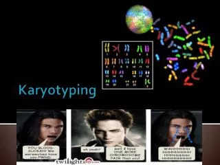
KARYOTYPING
KARYOTYPING. Dr. R.A. Siddique M.V.Sc PhD Scholar National Dairy Research Institute Karnal, (Haryana) 132001 India. CYTOGENETICS.

KARYOTYPING
E N D
Presentation Transcript
KARYOTYPING Dr. R.A. Siddique M.V.Sc PhD Scholar National Dairy Research Institute Karnal, (Haryana) 132001 India
CYTOGENETICS • Is the study of the structure and properties of chromosomes, chromosomal behaviour during mitosis and meiosis, chromosomal influence on the phenotype and the factors that cause chromosomal changes (Hare and Singh, 1979).
METHODOLOGY • Aseptic precautions • Preparation of RPMI 1640 medium • Collection of 10ml of blood with heparin • Setting of culture 8 ml of medium 0.1 ml of PHA-M 0.5 ml of blood/plasma 2 ml of autologus plasma/FCS • Incubate at 37C for 72 hours
METHODOLOGY… Harvesting of culture • Spindle inhibitors – Colchicine/colcemed (0.1g/ml) • Hypotonic treatment – 0.075M KCl • Fixation (3:1 methanol : acetic acid) • Preparation of slides • Slides stained with 4% Giemsa for 20-25min • Screening of slides to study the morphology of chromosome • Construction of karyotype
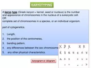
TERMS AND DEFINITIONS OF VARIOUS ABERRATIONS OF CHROMOSOMES • Ring( r) Minute (min) • Dicentric (d) Hyperdiploid (h) • Chromosome gap (sg) Chromatid deletion (td) • Fragment (f) Acentric fragment (af) • Translocation (t) Triradial (tr) • Quadriradial (qr) Pulverized chromosome (pu) • Pulverized chromosome (pu+) • Pulverized cell (puc) • Complex rearrangement (cr) • Polyploid (pp) or endoreduplication
BANDING OF CHROMOSOMES • G - Banding • Q - Banding • C - Banding • R - Banding • T - Banding • NOR - Banding • High Resolution Banding • Restriction Endonuclease Banding
Q-banding 1. Dehydrate the slides by dipping in alcohol with decreasing concentration 90%, 70% and 50% one min each. 2. Rinse in distilled water. . 3. Wash the slide in phosphate buffer at pH 6.8. 4. Stain the slide in quinacrine mustard (5 mg in 100 mI) or in quinacrine dihydrochloride 5% for 20 min. 5. Rinse in phosphate buffer and mount in the same buffer. 6. Examine under fluorescent microscope.
C-banding 1. Treat the slides in 0.2 N HCI for one hr at room temperature. 2. Rinse in de-ionized water. 3. Immerse in 1% barium hydroxide at 50°C for 5-15 min. 4. Rinse in deionized water. 5. Incubate at 60°C in 2XSSC buffer for one hr. 6. Rinse in de-ionized water and stain in 4% Giemsa stain for 90 min. 7. Rinse in de-ionized water, dry and examine under oil immersion.
R-banding 1. Age the slides for 7 -10 days . 2. Place the slides in a Coplinjar containing phosphate buffer ofpH 6.5 at 85°C and incubate for 20-25 min. 3. Stain the slides in 0.01% acridine orange in the phosphate buffer pH 6.5 for 4-6 min. Rinse in phosphate buffer and mount in the same buffer. 4. Examine under fluorescent microscope.
T -banding 1. Age the slide for 7 days. 2. Place.the slides in PBS pH 5.0 for 20-60 min at 87°C. 3. Rinse in PBS. 4. Stain in 3% Giemsa in phosphate buffer pH 6.8 at 87°C, leave for 5-30 min and rinse. 5. Slides are stained in Hoechst 33258 stain for 10 min (Hoechst stain 0.5 pg/m1 of phosphate buffer).Rinse in phosphate buffer and examine in fluorescent microscope. 6. Alternatively, the stained slides are covered with a cover slip and placed in a wet chamber under UV lamp for 2 to 3 hrs or under direct sunlight for 2 hrs. 7. Remove the cover slip and stain in Giemsa stain for 10 min. 8. Rinse in buffer, dry and mount in DPX.
METHODOLOGY G- Banding technique • Ageing of good slides for 10 days • Normal saline • Treated with trypsin 0.25% solution 10-15 sec • Immersed in 70% ethanol for few minutes • Stained with 10% Giemsa for 6-10min • Microphotograph good spreads • Construction of G-banded karyotype