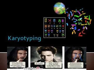
Karyotyping
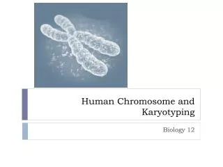
Karyotyping. What are Chromosomes. Chromosomes are structures found in the nucleus of cells Chromosomes carry all of our genes, and therefore all of our genetic information Humans have 46 chromosomes, or 23 pairs, to carry our approximately 25,000 genes

Karyotyping
E N D
Presentation Transcript
What are Chromosomes • Chromosomes are structures found in the nucleus of cells • Chromosomes carry all of our genes, and therefore all of our genetic information • Humans have 46 chromosomes, or 23 pairs, to carry our approximately 25,000 genes • The first 22 pairs are called autosomes • The 23rd pair are the sex chromosomes or gonosomesthis pair will either be XX or XY
Karyotyping • During mitosis, the 23 pairs of human chromosomes begin condensing during prophase, and reach their most condensed state during metaphase • A karyotype analysis usually involves blocking cells in mitosis and staining the condensed chromosomes with Giemsa dye. • The dye stains regions of chromosomes that are rich in the base pairs Adenine (A) and Thymine (T) producing a dark band
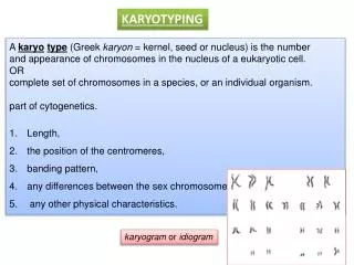
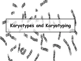
Karyotypes • A karyotype is a pattern or picture of chromosomes. The chromosomes are paired and arranged according to size • Each chromosome is paired with its homologous chromosome • its exact match in size and structure, though the homologous chromosomes may carry different alleles of the same gene • Then the chromosome pairs are labeled • The autosomes are numbered 1 through 22 according to size • The sex chromosomes are a different story • The X and X or the X and Y are paired, then placed at the end, even though they are not necessarily the smallest chromosomes • These chromosomes do not receive a number - just XX or XY
Human Karyotyping • A karyotype is an actual photograph of the chromosomes from one cell. • Karyotypes are usually done using blood cells, fetal skin cells (from amniotic fluid or the placenta) and occasionally bone marrow cells • It takes at least one week to complete a karyotype
Clinical Steps to Karyotyping 1. Sample Collection 2. Separating cells 3. Growing Cells 4. Synchronizing Cells 5. Releasing Chromosomes from their cells 6. Staining Chromosomes 7. Analysis a. Counting & Sorting of chromosomes b. Looking at Chromosomes Structure
Sample Collection • In newborns, a blood sample which contains red bloods cells, white blood cells, serum and other fluids is collected. • A karyotype will be done on the white blood cells which are actively dividing (a state known as mitosis).
How do we get blood samples from a fetus • During pregnancy, the sample can either be amniotic fluid collected during an amniocentesis • The amniotic fluid contains fetal skin cells which are used to generate a karyotype • or a piece of the placenta collected during a chorionic villi sampling test (CVS). • The important thing is to choose a cell type that is actively dividing, some cells, like nerve cells, divide infrequently or never!
Separating the Cells • In order to analyze chromosomes, the sample must contain cells that are actively dividing (or in mitosis). • In blood, the white blood cells are actively dividing cells. • Sometimes adult cells are treated with chemicals such as phytohemaglutenin (PHA) to induce mitotis • Most fetal cells are actively dividing. • Once the sample reaches the cytogenetics lab, the non-divided cells are separated from the dividing cells using special chemicals.
Growing Cells • In order to have enough cells to analyze, the dividing cells are grown in special media or a cell culture. • This media contains chemicals and hormones that enable the cells to divide and multiply. • This process of “culturing” the cells can take 3 to 4 days for blood cells, and up to a week for fetal cells
Synchronizing Cells • The best mitotic phase for chromosome analysis is prometaphase or metaphase • In order to get all the cells to this specific stage of cell division, the cells are treated with a chemical which stops cell division at the point where the chromosomes are the most compact. • cholicine
Releasing Chromosomes From Their Cells • In order to see these compact chromosomes under a microscope, the chromosomes have to be out of the white blood cells. • This is done by treating the white blood cells with a hypotonic solution that causes • Chromosomes to spread apart for easier viewing • And causes cells to burst • This is done while the cells are on a microscopic slide. The leftover debris from the white blood cells is washed away, and the chromosomes are fixed to the slide.
Staining the Chromosomes • Chromosomes are naturally colorless. • In order to be able to tell one chromosome from another, a special dye called Giemsa dye is applied to the chromosomes on the slide. • Giemsa dye stains regions of chromosomes that are rich in the bases adenine (A) and thymine (T). • When stained, the chromosomes look like strings with light and dark bands. • Each chromosome has a specific pattern of light and dark bands which enables cytogeneticist to tell one chromosome from another.
G Banding • 1st use a protease to digest any protins • Giemsa reagent consists of a mixture od dyes including • Aminophenothiazine dyes(basic) • Eosin (acidic) • Produces light & dark bands • Patterns of banding change with the stage of mitosis • There may be as many as 1,000 bands in early prophase, and as few as 300 on a given chromosome in metaphase
G Banding Light Bands Dark Bandss AT rich Highly coiled regions of DNA Contains few genes Most genes in these regions are tissue specific, and so not transcribed in all cells • GC rich • Most genes are in light bands • Light bands represent relatively open (uncoiled) regions of DNA making transcription easier
Analysis • Once chromosomes are stained, the slide is put under the microscope and the analysis of the chromosomes begins. • A picture is taken of the chromosomes and at the end of the analysis • This is known as a “metaphase spread” • Take a picture of the spread using a special microscope with attached camera
Critical Thinking! • When we draw chromosomes, we typically draw them in the shape of the letter X • What does this represent (ie, label the important parts of the X structure) • Why, in the above metaphase spread are the chromosomes NOT in the X shape, but rather linear? • Hint: remember, there are 2 types of cell division
Analysis: Step 1 • The first step of the analysis is counting the chromosomes. • Most humans have 46 chromosomes. • People with Down syndrome have 47 chromosomes. • It is also possible for people to have missing chromosomes or more than one extra chromosome. • By looking at just the number of chromosomes, it is possible to diagnose different conditions including Down syndrome, Turners, Kleinfelters
Analysis: Step 2 • Sort the chromosomes by • comparing chromosome length, • the placement of centromeres (the areas where the two chromatids are joined), • the location and sizes of G-bands. • The chromosomes pairs are numbered from largest (number 1) to smallest (number 22). • There are 22 pairs of chromosomes, called autosomes, which match up exactly. • 2 sex chromosomes • two X‘s is a female • X and a Y is a male.
Analysis: Step 3 • In addition to looking at the total number of chromosomes and the sex chromosomes, the cytogeneticist will also look at the structure of the specific chromosomes to make sure that there is no missing or additional material, no structural abnormalities like • Translocations • Deletions • Duplications
What we will do • Unfortunately in high school we aren’t allowed to stick needles in our students and/or work with human bodily fluids • Possible spread of infectious diseas • Parents complain • We will practice karyotyping from the analysis standpoint
3 Criteria used to Identify a Chromosome • The analysis involves comparing chromosomes for their • Length • placement of centromeres (areas where the two chromatids are joined) • location and sizes of G-bands.
Typical Chromosome Structure • Each chromosome has 2 arms separated by a constriction point called the centromere • Metacentric: term used to describe chromosomes with centromere at or near the center of the chromosome • Submetacentric: term used to describe chromosomes with centromere slightly displaced from the center
Typical Chromosome Structure • Very submetacentric: term used to describe chromosomes with centromere halfway between the middle and the tip of the chromosome • Acrocentric: term used to describe chromosomes with centromere very near one end
Typical Chromosome Structure • Telocentric: term used to describe chromosomes with centromere at the very tip of one end • These are absent in human chromosomes
Typical Chromosome Structure • Notice, the centromere divides the chromosome into 2 separate regions • In general • The shorter chromosome arm is designated p for petite • The longer arm is designated the q arm, because the letter q comes after the letter p in the alphabet
How To Start • Open your worksheet titled Key to Human Chromosomes • It is a dichotomous key • Start by cutting chromosomes out of your metaphase spread • Divide all chromosomes into one of the 2 class sizes • Class 1 = 1st 12 chromosome pairs (24), and X chromosomes • So class 1 will contain 25 chromosomes for a boy and 26 chromosomes for a girl • Class 2 = the rest of the smaller chromosomes
Nomenclature • Normal female 46, XX • Normal Male 46, XY • Telomeres, centromeres, and a number of prominent bands areused as landmarks on a chromosome
Nomenclature • A section of a chromosome between 2 landmarks is called a region • Regions are numbered 1,2,3 in both directions starting with the centromere • Bands within a region are numbered according to the same rule • Example: the 1st band in the 2nd region of the p arm of chromosome #1 is indicated as follows • 1p21
So what does this mean: 14q32 • 14 refers to chromosome # 14 • q refers to the q arm (the longer arm) of chromosome 14 • 3 refers to region 3 of the q arm of chromosome 14 • And 2 refers to the 2nd band in the 3rd region of the q arm in chromosome 14
Nomenclature • There may also be sub-bands • Decimal points are used to indicate what sub-band • Example: 14q32.3 • Refers to the 3rd (last) sub-band within the 2nd band of region 3 in the q arm of chromosome 14
Numerical Chromosome Abberations for Sex Chromosomes • 45, X 45 chromosomes, 1 X chromosome • 47, XXY 45 Chromosomes, two X chromsomes one Y chromosome
Numerical Chromosome Abberations • Use a + or – sign • before the symbol to indicate additional or missing whole chromosomes • After a symbol to indicate additional or missing segments of a chromosome • 47, XY, +21 an additional 21st chromosome • 46,XX, 1q+ female karyotype with 46 chromosomes showing an increase in the length of the long arm of chromosome 1
Nomenclature • Question marks are used to indicate uncertainty • 45, XY, -?8 • 45 chromosomes • XY is male • Missing a chromosome, probably #8 • There is special nomenclature for inversions, deletions, translocations, insertions, etc.; but its is beyond the scope of this class
Other Types of Staining • C-Banding: stains constitutive heterochromatin • Regions of chromosomes that are highly compacted containing highly repetitive DNA • Results from selective removal of the chromosomes except from regions of the C-Bands • Used for determining presence or absence of a centromere, as well as for studying abnormalities of the centromeric region
Other Types of Staining • Silver Staining for NOR • Ribosomal DNA, which codes for ribosomal RNA, occurs in multiple copies on the short arms of all acrocentric chromosomes • Silver stain results in the deposition of silver grains on the active nucleolar organizer regions (NORs) • In other words, NORs which were actively transcribing their rRNA’s in the preceding interphase, contain the proteins to which thre silver grains bind
FISH • Fluorescence In Situ Hybridization • Involves precise annealing of ssDNA probes to complementary target sequences • Can be visualized with a fluorescent microscope by using a probe directly labeled with a fluorophore • Can be used to find • If a specific gene is present (and or duplicated) • And if it is in the correct region of the correct chromosome
WCP • Whole Chromosome Painting • Same idea as FISH, but you use a mixture of many fluorophore labeled probes • Must also use unlabeled blocking DNA to supress highly repetitive regions common to many different chromosomes • Allows for • Chromosome enumeration • Identification of chromosomal structural rearrangements
WCP • Particularly useful in solid tumor and linical genetic research because it has the ability to aid in the analysis of complex and cryptic translocations • Also helpful in research that is evaluating the extent of chromosomal breakage and translocations following exposure to mutagenic chemicals or radiation
Using A Karyotype • A karyotype allows a cyto-geneticist or lab technician to examine the chromosomes and see if there is anything extra or missing, or if the structure of the chromosomes is grossly different than usual
Trisomy 21 • Trisomy 21 is the presence of 3 chromosome 21’s. • Trisomy 21 causes the condition commonly known as Down syndrome • The extra chromosome leads to the specific characteristics of Down syndrome, some of which are very familiar • not all individuals with Down syndrome will show the exact same characteristics there is a great deal of variability